
|
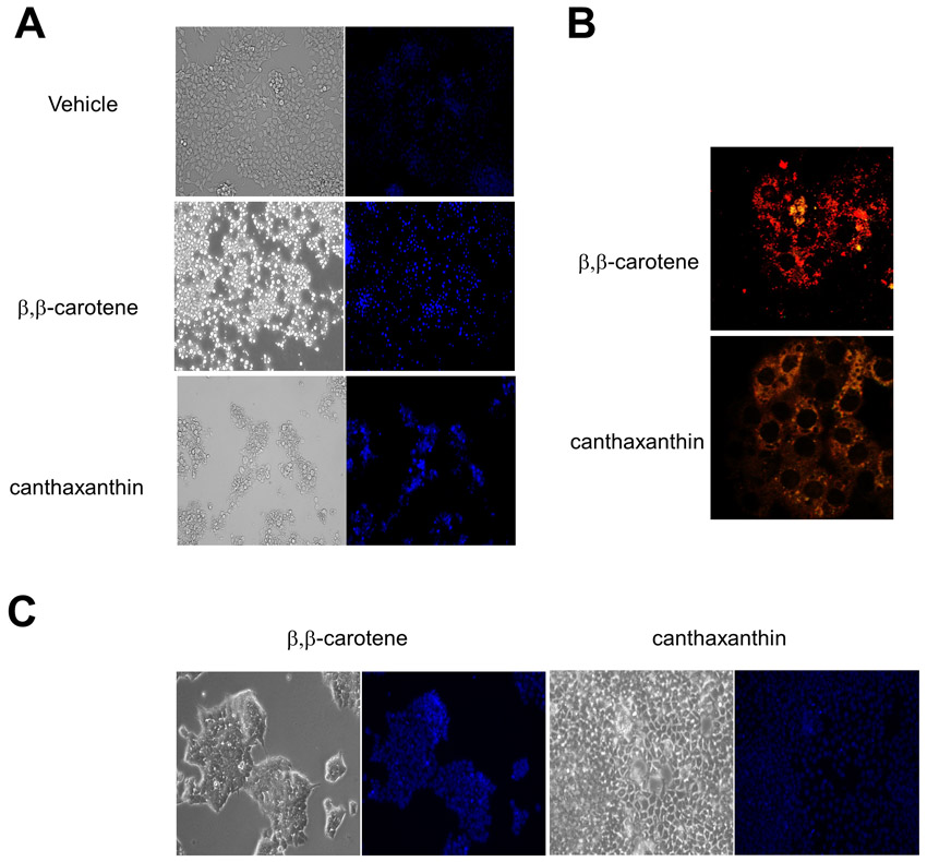
Fig. S4 Effects of recombinant Bcdo2 expression in HepG2 and siRNA knockdown of Bcdo2 in T47D cells. (A) HepG2 cells were incubated in the presence of vehicle, 1 μM β,β-carotene and 1 µM canthaxanthin, respectively. After incubation with Hoechst 33342 dye, cells were examined for chromatin condensation under a fluorescence microscopic system at 20× magnification. (B) BCDO2 was expressed as a recombinant protein in HepG2 cells. Forty to 48 hours post transfection, cells were incubated in the presence of 1 μM β,β-carotene and 1 μM canthaxanthin, respectively. After 2 hours, the mitochondrial membrane potential was assessed by using the JC-1 stain. Cells were imaged at 20× magnification under a confocal microscope. Red fluorescence of JC-1 aggregates is indicative for intact mitochondria. Green fluorescence of JC-1 monomers dispersed in the cytoplasm indicates mitochondria with depolarized membrane potentials. (C) T47D cells were pre-treated with siRNA specific to human Bcdo2 for 72 hours. Then, cells were incubated with 1 μM β,β-carotene or 1 7mu;M canthaxanthin. After incubation with Hoechst 33342 dye, cells were examined for chromatin condensation under a fluorescence microscopic system at 20× magnification