
|
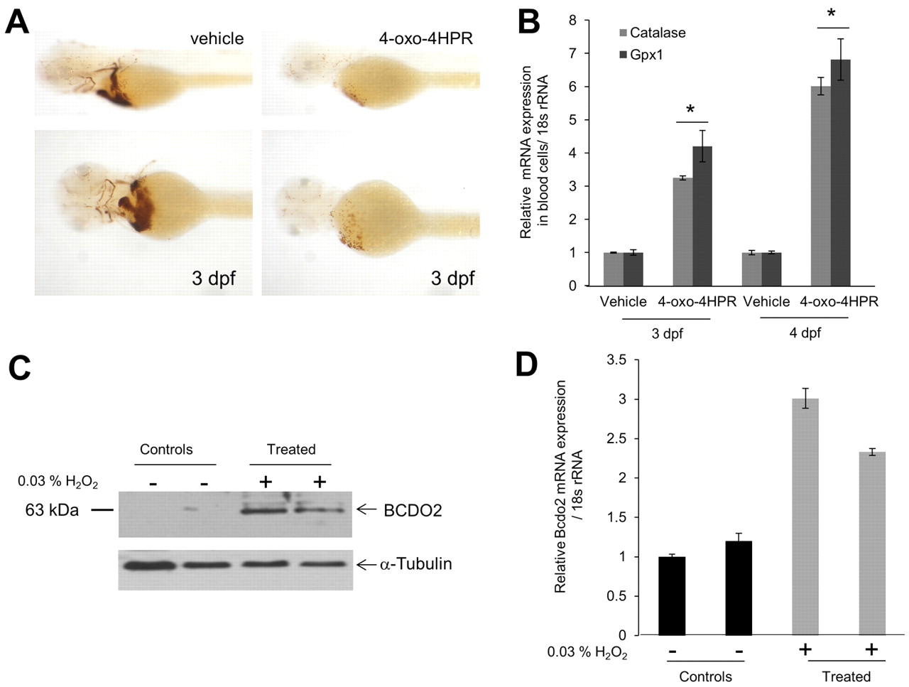
Fig. 6 4-oxo-fenretinide (4-oxo-4HPR) induces apoptosis of blood cells and oxidative stress in zebrafish larvae. All embryos were raised in the presence of 200 μM 1-phenyl-2-thiourea (PTU) to inhibit pigmentation at 28°C. Two days post-fertilization, embryos (n=50 for each condition and experiment) were manually dechorionized and incubated in egg water containing 1 μM 4-oxo-fenretinide (4-oxo-4HPR) or vehicle only [0.1% (v/v) DMSO]. (A) After 24 hours, hemoglobin staining was performed using the o-Dianisidine dye. Representative images were taken under a Leica stereo microscope at 1.6× (top panels) and 4.0× (bottom panels) magnifications. (B) RNA from isolated blood cells was assessed for glutathione peroxidase 1 (gpx1) and catalase mRNA expression. The 18s rRNA probe set was used as the endogenous control. *P<0.05 compared with controls. Data are mean±s.e.m. (C,D) Larvae (2 dpf) were incubated in the presence or absence of 0.03% hydrogen peroxide (H2O2) for 24 hours. (C) Protein extracts from 12 larvae per lane were subjected to immunoblot analysis for BCDO2. α-Tubulin was used as protein loading control. (D) Total RNA was isolated from 12 larvae and bcdo2 expression was determined by qRT-PCR. Data are mean±s.e.m.