
|
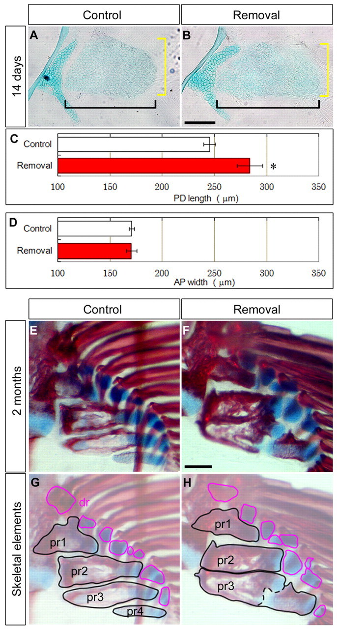
Fig. 6
Repeated AF removal affected endoskeletal bones. (A,B) Alcian Blue staining of the pectoral fin 14 days after AF removal (three times). Scale bar: 100 μm. (C,D) PD length (C, black brackets in A,B) and AP width (D, yellow brackets in A,B) of endoskeletal disc after removing the AF three times. Error bars indicate s.e.m. Data were analyzed by Student’s t-test (*P<0.05). Measurements are shown in supplementary material Table S2. (E-H) Alcian Blue and Alizarin Red staining of the pectoral fin 2 months after AF removal. Specimens (E,F) are outlined and highlighted, focusing on endoskeletal bones (G,H). Proximal radials (pr) are outlined in black. Distal radials (dr) are outlined in red. Scale bar: 100 μm.