
|
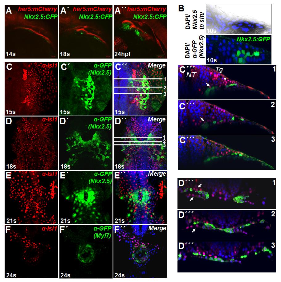
Fig. S5
(related to Figure 5). Isl1 expression in cardiac progenitor cells.
(A, A′, A′′) Expression of the Nkx2.5:GFP transgene in anterior lateral plate mesoderm, adjacent to pharyngeal endoderm, marked by her5:mCherry (Tallafuss and Bally-Cuif, 2003) at different developmental stages. (B) In situ hybridization for Nkx2.5 and immunostaining for GFP (Nkx2.5), showing a similar domain of expression of the Nkx2.5:GFP transgene and the Nkx2.5 mRNA (C, C′, C′′, C′′′). Confocal images of Nkx2.5:GFP transgenic embryos stained with anti-Isl1 and anti-GFP antibodies at the 15 somite stage. C′′ shows the position of the transverse (xz) optical sections presented in C′′′1, 2, 3. (D, D′, D′′, D′′′) Confocal images of Nkx2.5:GFP transgenic embryos stained with anti-Isl1 and anti-GFP antibodies at the 18 somite stage. D′′ shows the position of the transverse optical sections presented in D′′′1, 2, 3. (E, E′, E′′) Confocal images of Nkx2.5:GFP transgenic embryos stained with anti-Isl1 and anti-GFP antibodies at the 21 somite stage. (F, F′, F′′) Confocal images of Tg(myl7:EGFP-HsHRAS)s883 embryos stained with anti-Isl1 and anti-GFP antibodies at the 24 somite stage. NT, neural tube, Tg, trigeminal placode.
Reprinted from Developmental Cell, 23(1), Witzel, H.R., Jungblut, B., Choe, C.P., Crump, J.G., Braun, T., and Dobreva, G., The LIM Protein Ajuba Restricts the Second Heart Field Progenitor Pool by Regulating Isl1 Activity, 58-70, Copyright (2012) with permission from Elsevier. Full text @ Dev. Cell