
|
Fig. S2
(related to Figure 3). Confirmation of the efficiency and the specificity of morpholino targeting
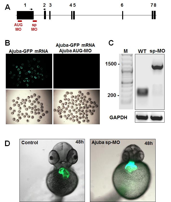
(A) Schematic representation of the Ajuba genomic locus and the binding sites of the AUG and the splice morpholino (shown in red). Arrows indicate the positions of the primer pair used for the RT-PCR analysis in (C), (B) To confirm the efficiency of the AUG morpholino we constructed a pCS2-Ajuba-GFP plasmid, in which the binding sequence of the AUG morpholino was fused in frame with the GFP cDNA. Embryos injected with 200 ng Ajuba-GFP mRNA, synthesized from the pCS2-Ajuba-GFP plasmid, showed GFP expression. In contrast in embryos injected with 200 ng Ajuba-GFP mRNA and 5 ng Ajuba AUG MO, GFP fluorescence was not observed. (C) The efficiency of Ajuba sp-MO was analyzed by PCR using primers spanning the first intron of the Ajuba gene. This analysis indicated loss of spliced Ajuba mRNA in embryos injected with the Ajuba sp-MO. (D) Control or Ajuba sp-MO injected Tg(myl7:EGFP-HsHRAS)s883 embryos at 48 hpf. Frontal views, showing an enlarged atrium and reversed left–right asymmetry of the heart.
Reprinted from Developmental Cell, 23(1), Witzel, H.R., Jungblut, B., Choe, C.P., Crump, J.G., Braun, T., and Dobreva, G., The LIM Protein Ajuba Restricts the Second Heart Field Progenitor Pool by Regulating Isl1 Activity, 58-70, Copyright (2012) with permission from Elsevier. Full text @ Dev. Cell