
|
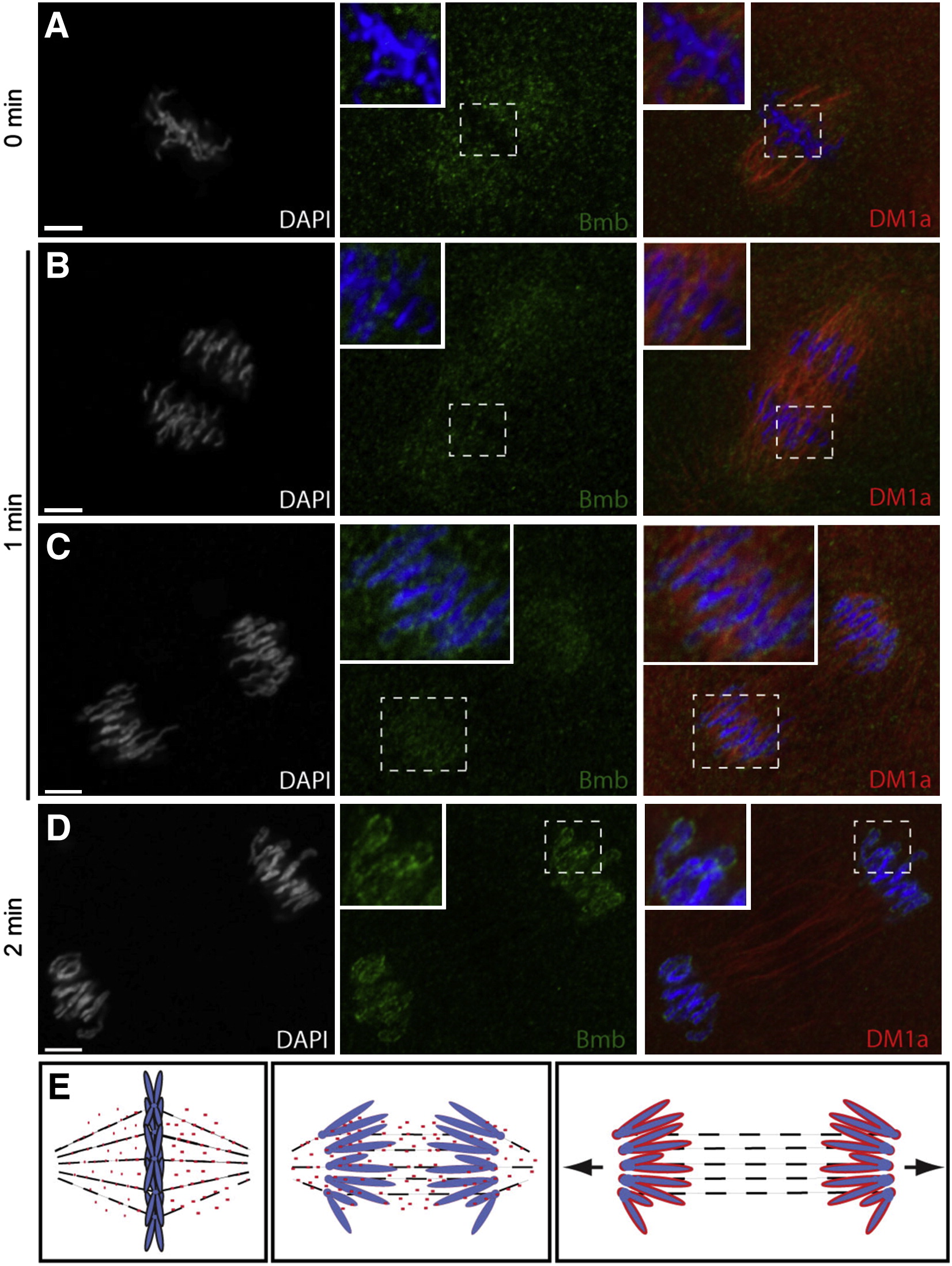
Fig. 4
Bmb Protein Dynamics during Mitosis(A) Bmb protein localizes as distinct foci to the mitotic spindle region during metaphase (n = 4).(B) During anaphase, Bmb is interspersed between the separating chromosomes (inset; n = 3) and in the region of the mitotic spindle.(C) Later in anaphase, Bmb foci are decorating the chromosomes (inset; n = 3).(D) At the 2 min time point, Bmb surrounds the individual chromosomes, which are still separating (n = 5).(E) Schematic summarizing Bmb localization during metaphase to anaphase/telophase. Arrows in the right panel indicate that the karyomeres will continue to move to their final central position in the cell. 0 min corresponds to metaphase at the 32- to 64-cell transition. Bmb (green), microtubules (DM1a, red), and chromosomes (DAPI, white and blue in merge).Scale bar, 5 μm, and insets show 2× enlargements. See also Figure S2.
Reprinted from Cell, 150(3), Abrams, E.W., Zhang, H., Marlow, F.L., Kapp, L., Lu, S., and Mullins, M.C., Dynamic Assembly of Brambleberry Mediates Nuclear Envelope Fusion during Early Development, 521-532, Copyright (2012) with permission from Elsevier. Full text @ Cell