
|
Fig. S2
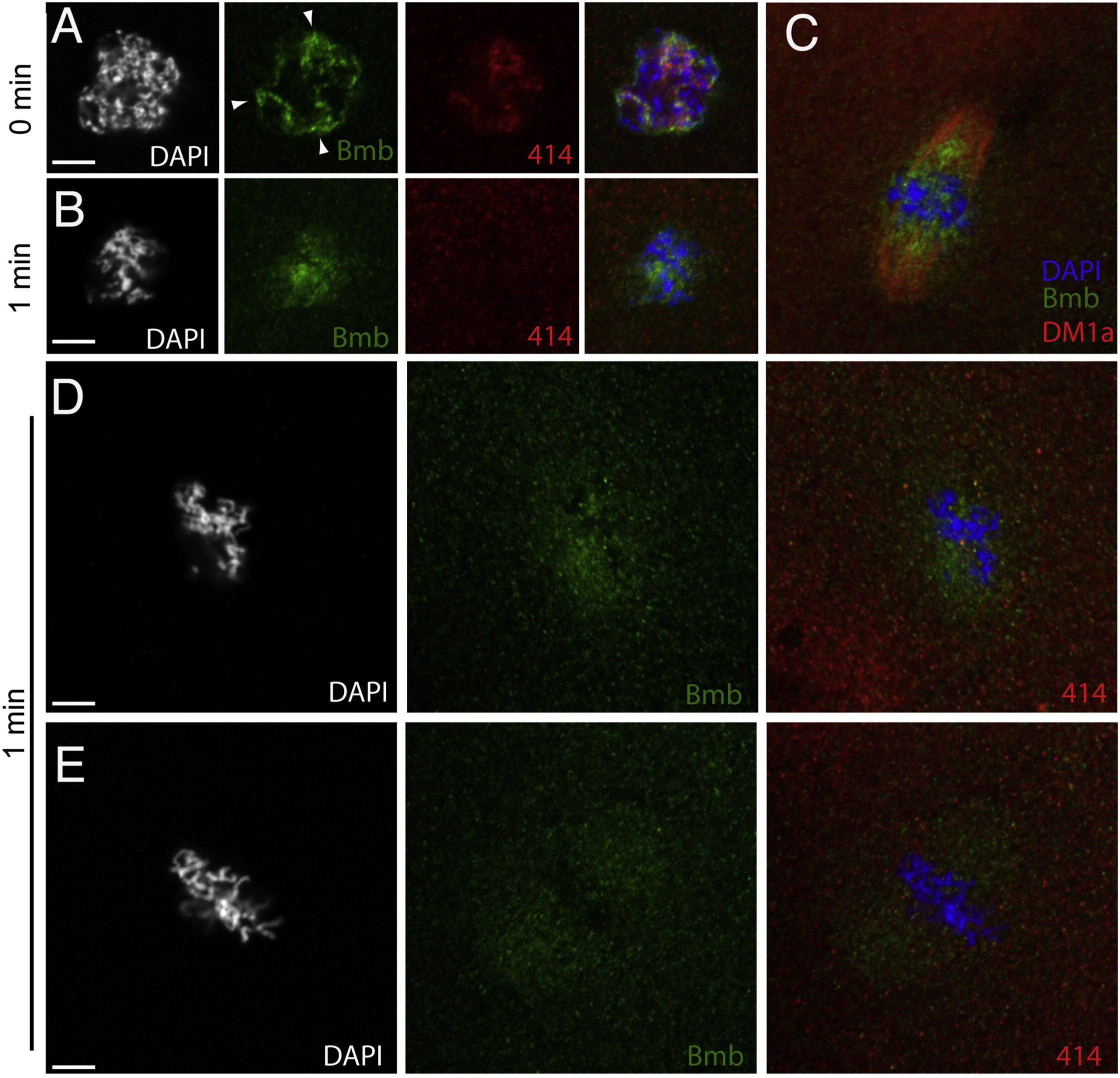
Bmb Localization at Prophase through Metaphase, Related to Figure 4(A) Embryos fixed at one-minute time points were stained with anti-Bmb (green), mab414 (red) and DAPI (white/blue). During prophase (A) (0 min), Bmb signal can be seen concentrated in regions of the nucleus as the chromatin condenses (n = 5). (B) At prometaphase (B) (1 min), concentrated Bmb begins to disperse (n = 5). (C) (C) is an analogous time point to (B) taken from an independent experiment staining for Bmb (green) and microtubules (red, DM1a) indicating that Bmb protein becomes more concentrated in the region of the spindle closer to the chromosomes (n = 5). (D) A different sample from the same time point (1 min) reveals Bmb signal becoming more dispersed in a punctate arrangement. At this point as in (B) mab414 is completely dispersed (n = 3). (E) Once the chromosomes are aligned at the metaphase plate, Bmb becomes more evenly distributed within the spindle region (n = 5). 0 min corresponds to early prophase at the 32-to-64 cell transition. Images correspond to Z-projections of individual confocal Z-planes.
Reprinted from Cell, 150(3), Abrams, E.W., Zhang, H., Marlow, F.L., Kapp, L., Lu, S., and Mullins, M.C., Dynamic Assembly of Brambleberry Mediates Nuclear Envelope Fusion during Early Development, 521-532, Copyright (2012) with permission from Elsevier. Full text @ Cell