
|
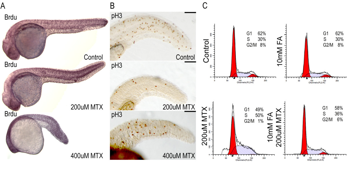
Fig. 6 Abnormal cell cycle properties of methotrexate treated embryos A) After 24 hours of 200 μM or 400 μM methotrexate exposure, embryos are capable of incorporating BrdU via DNA synthesis. B) Immunohistochemistry for the mitotic marker phospho-histone H3 shows a decreased number of mitotic cells in methotrexate treated embryos. C) However, FACS analysis clearly indicates methotrexate treated embryos do not display WT cell cycling profiles, with a probable S-phase accumulation. Co-treatment with folinic acid and methotrexate restores cell cycle profiles. Lateral views of 24 hpf embryos. Scale bar: 100 μm.