
|
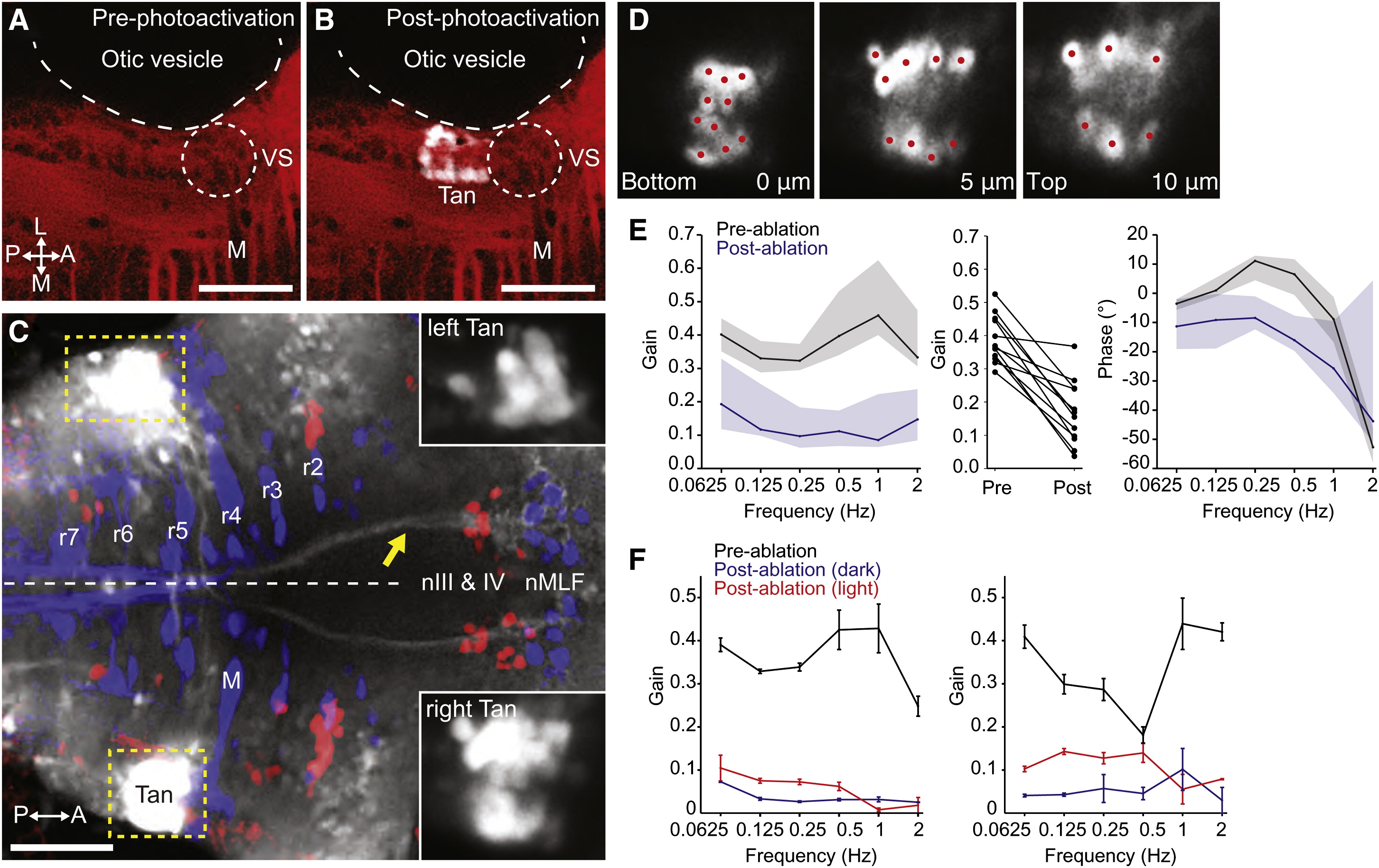
Fig. 5 Tangential Nucleus Neurons Are Required for the VOR(A–C) Labeling of the tangential nucleus with C3PA-GFP in a lyn-mCherry background.(A) Lyn-mCherry expression (red) revealed the location of the Mauthner cell (M) and vestibulo-spinal neurons (VS), which served as landmarks allowing us to target the tangential nucleus (Tan) in r5.(B) After photoactivation, a cluster of cells in the left Tan nucleus is visible (white).(C) Axonal projections to nIII, nIV, and nMLF were visible after bilateral labeling of tangential nuclei. The yellow arrow indicates axons ascending in the left MLF, derived from right-sided photoactivated Tan neurons. Insets show the photoactivated tangential nuclei, with dynamic range adjusted to reveal the cell bodies.(D) Three optical sections through the right tangential nucleus of the same larva shown in (C). Photolabeled neurons were individually ablated by spiral-scanning a pulsed infrared laser beam that was centered on the somata of each neuron in turn (red spots).(E) Laser ablation of C3PA-GFP-labeled tangential neurons caused a substantial reduction in the gain of the VOR. Left panel shows Bode plot of gain, presented as median ± interquartile range (n = 12). Middle panel shows change in mean gain across frequencies for all 12 larvae. Right panel shows Bode plot of phase.(F) Data from two fish that displayed the greatest reduction in gain after tangential ablation. Data shown as mean ± SEM. Torsional eye rotations could be evoked by visual input at low frequencies.All images are dorsal views, anterior right. Scale bars represent 50 μm. See also Figure S5.