
|
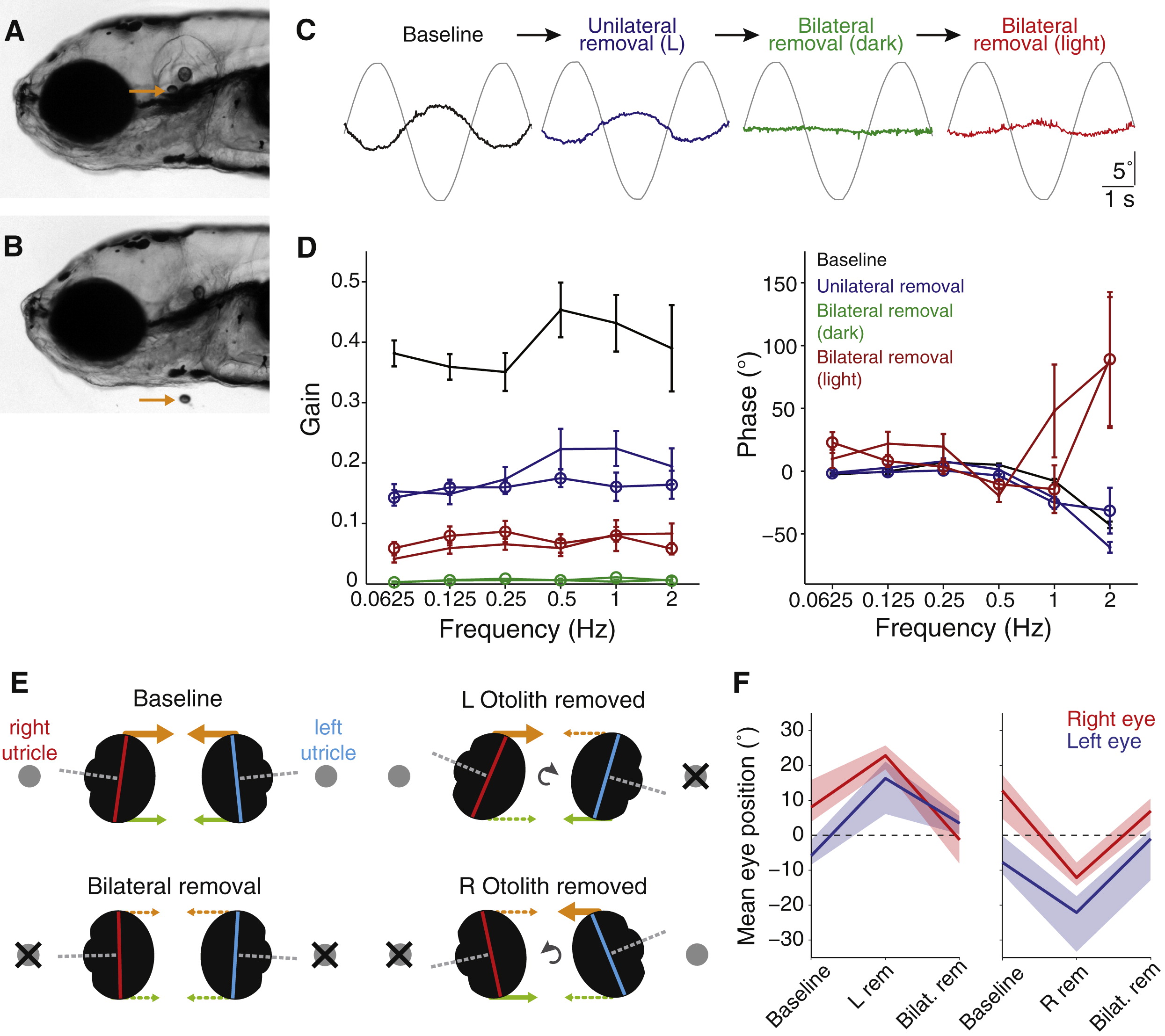
Fig. 3 Otolith Removal(A) Lateral view of a 5 dpf larva (anterior left). Arrow points to the utricular otolith.(B) Surgical removal of the utricular otolith. Note the collapse of the otic vesicle after surgery.(C) Example traces from a representative fish (left eye), showing torsional eye movement responses to a 0.25 Hz sinusoidal stimulus after sequential perturbations: baseline performance before surgery, after removal of only the left otolith, after both otoliths are removed, and after both are removed and tilt stimuli were delivered in the light (in all other cases fish were tested in the dark).(D) Bode plots of gain and phase for utricle removal experiments. Lines with open symbols represent right eye data, lines without represent left eye data. We combined data for experiments in which the left otolith was removed first (n = 6) and in which the right otolith was removed first (n = 4) because for both eyes, removal of either otolith had similar effects. Data are shown as mean ± SEM. Phase data are not shown for the “Bilateral removal (dark)” condition because this parameter can not be accurately estimated when gain approaches zero.(E) Schematic representation of the vertical eye positions that were measured under different experimental conditions. By viewing the larvae head-on, the long axes of the right (red) and left (blue) eyes were measured to determine vertical eye position. Orthogonal visual axes are shown in gray, the predicted superior extraocular muscle forces are shown in orange, and inferior muscle forces in green.(F) Summary vertical eye position data, presented as median ± interquartile range for experiments in which the left utricular otolith was removed first (left, n = 10) or the right otolith was removed first (right, n = 4).See also Figure S3 and Movies S1 and S2.