
Fig. S2
- ID
- ZDB-IMAGE-120803-8
- Publication
- Talbot et al., 2012 - fras1 shapes endodermal pouch 1 and stabilizes zebrafish pharyngeal skeletal development
- All Figures
- Figures for Talbot et al., 2012
|
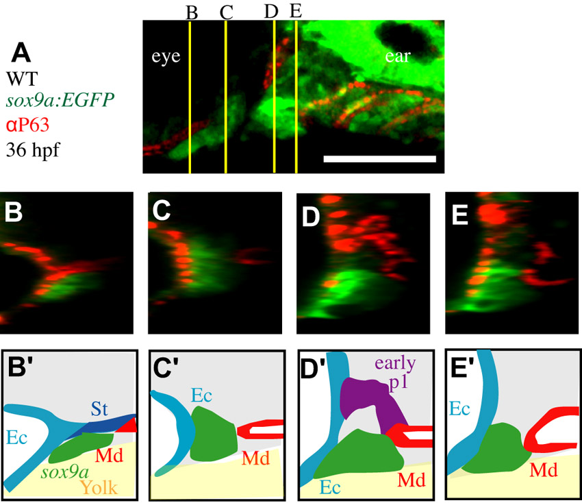
Fig. S2 Early pouching appears normal in fras1 mutants. (A) Confocal section of WT pharyngeal arches, with pre-cartilage cells labeled using sox9a:EGFP and epithelial nuclei labeled using anti-P63; shown anterior to left, dorsal up. Yellow lines indicate section levels for B-E. (B-E) Four confocal transverse sections through arches one and two, reconstructed from a confocal stack of the embryo imaged for A, shown lateral to the left, dorsal up. (B′-E′) Epithelial morphology. At 36 hpf a tube of endoderm lies medial to the length of arches; early-p1 separates arch one and two mesenchyme dorsally, and posterior pouches fully separate posterior arch mesenchyme. Ec, surface ectoderm; Md, medial endoderm; p2, pouch 2; p3, pouch 3; p4, pouch 4; St, stomadeum. Scale bar: 100 μm.