
Fig. 5
- ID
- ZDB-IMAGE-120803-5
- Genes
- Publication
- Talbot et al., 2012 - fras1 shapes endodermal pouch 1 and stabilizes zebrafish pharyngeal skeletal development
- All Figures
- Figures for Talbot et al., 2012
|
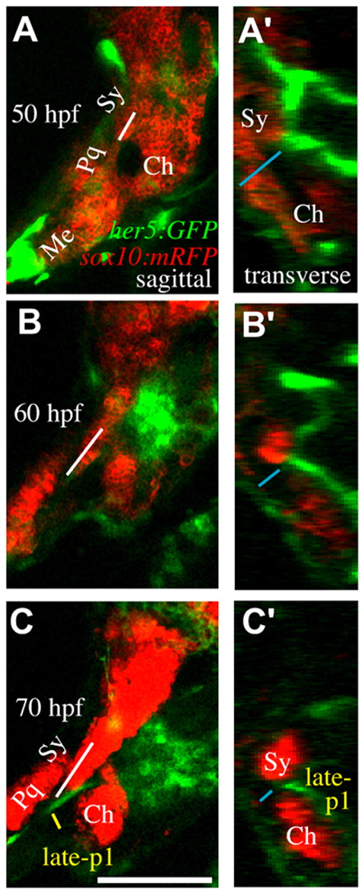
Fig. 5 Time-lapse microscopy reveals concurrent late-p1 and symplectic outgrowth. (A-C′) sox10:mRFP labels cartilages and her5:GFP labels late-p1 and some neural cells in these sections. Still images from time-lapse microscopy showing symplectic growth (white line in A-C) and endoderm-ectoderm distance (blue line in A′-C′). (A-C) Sagittal confocal section, anterior to left, dorsal up. By 70 hpf, symplectic tip extends out of the plane of section. (A′-C′) Transverse view, lateral to left, dorsal up, constructed from the same confocal stacks. Between 50 and 70 hpf, late-p1 moves laterally through a region between the symplectic and ceratohyal cartilages. By 70 hpf, development is slightly delayed, resulting in a mild reduction of late-p1 and cartilage formation. Scale bar: 100 μm.