
Fig. 1
- ID
- ZDB-IMAGE-120803-1
- Genes
- Antibodies
- Publication
- Talbot et al., 2012 - fras1 shapes endodermal pouch 1 and stabilizes zebrafish pharyngeal skeletal development
- All Figures
- Figures for Talbot et al., 2012
|
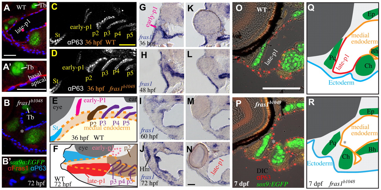
Fig. 1 Zebrafish fras1 mutants show specific late-p1 defects. (A-B′) Transverse sections of antibody-stained tissues, oriented lateral to left, dorsal up. (B) At the section level of late-p1, fras1 mutant endoderm does not extend as far laterally (asterisk) as WT endoderm at 72 hpf. (A′,B′) High-resolution detail near trabecula (Tb) from A and B shows that (A′) WT Fras1 is deposited basal to the epithelial nuclei marker and (B2) anti-Fras1 label is absent from fras1 mutants. Because these insets are from the roof of the mouth, basal is up and lateral is to the left. (C-E) By contrast, early pouches appear normal in fras1 mutants. (C,D) Confocal section of (C) WT and (D) fras1 mutant epithelia at 36 hpf, oriented anterior to left, dorsal up. Note that the early forming portion of pouch 1 is only mildly misshapen in fras1 mutants and posterior pouches appear normal. (E) Diagram of WT pouch structure at 36 hpf, viewed with anterior to left, dorsal up. Medial endoderm (orange dashed lines) lies beneath the plane of section shown in G and H (see supplementary material Fig. S2). Yolk is in yellow and somatic tissue is gray. (F) Diagram of pouch derivatives at 72 hpf, viewed with anterior to left, dorsal up. Between 36 and 72 hpf, late-p1 has protruded laterally, early-p1 has been covered by cartilages, and pouch 2 (p2) has extended posteriorly, covering posterior pouches. (G-N) RNA in situ hybridization on tissue sections, shown lateral to left, dorsal up. Epithelial fras1 expression lines pharyngeal arches during late-p1 formation. (G-J) Sections through early-p1 show that this early forming pouch is eventually covered by the hyomandibular cartilage, whereas (K-N) late-p1 protrudes laterally, anterior to early-p1. (O,P) Transverse sections of antibody-stained tissues, oriented lateral to left, dorsal up. Endodermal pouching defect persists in fras1 mutants at 7 dpf (asterisk). (Q,R) Illustrations of 7-dpf tissue sections, indicating the location of late-p1. Endoderm abbreviations: early-p1, early forming portion of pouch 1; late-p1, late-forming portion of pouch 1; p3, pouch 3; p4, pouch 4; p5, pouch 5; St, stomadeum. Cartilage abbreviations: Pq, palatoquadrate; Ch, ceratohyal; Bh, basihyal; Ep, ethmoid plate; Hm, hyomandibular. Scale bars: 50 μm (in A,A2,C,O for B,B2,D,P, respectively; in N for G-N).