
|
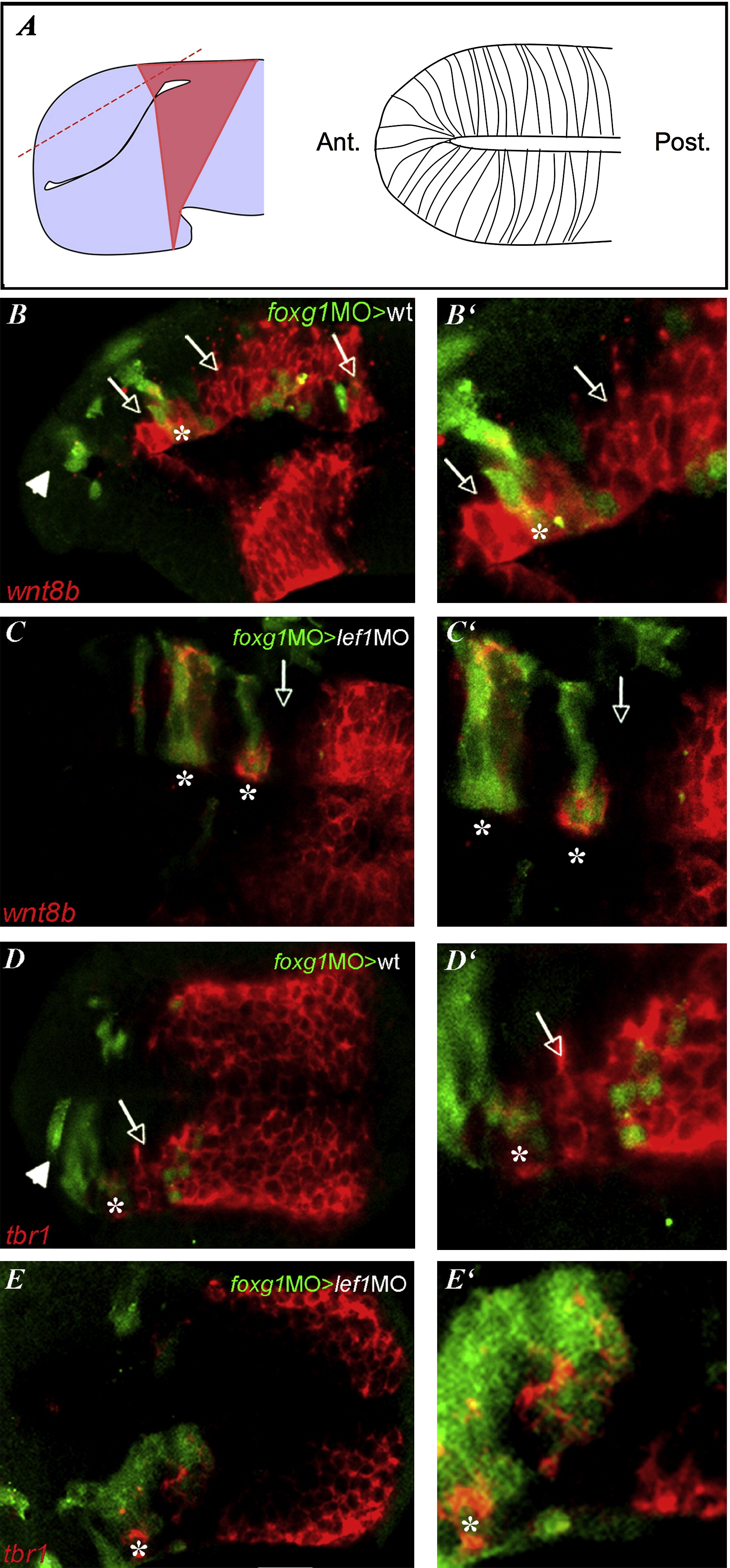
Fig. 6 Foxg1 Loss of Function Acts Cell Autonomously and Nonautonomously for the Restriction of Pallial Identity(A–E′) All embryos are at the 20ss. All panels show telencephalic dorsal views; anterior is oriented toward the left. The right column shows high magnification of the images on the left. (A) Schematic explaining the plan of section of the confocal images below. The normal wnt8b expression domain is pale red. (B–E) Confocal images of mosaic embryos with transplanted foxg1MO cells in green and (B–C′) wnt8b or (D–E′) tbr1 expression in red. Asterisks indicate foxg1MO cells, and arrows show neighboring host cells. Host embryos are (B, B′, D, and D′) WT or (C, C′, E, and E′) lef1 morphants. The arrowhead in (B) and (D) indicates the foxg1MO cells located in the ventral telencephalon.
Reprinted from Developmental Cell, 16(4), Danesin, C., Peres, J.N., Johansson, M., Snowden, V., Cording, A., Papalopulu, N., and Houart, C., Integration of telencephalic Wnt and hedgehog signaling center activities by Foxg1, 576-587, Copyright (2009) with permission from Elsevier. Full text @ Dev. Cell