
Image
|
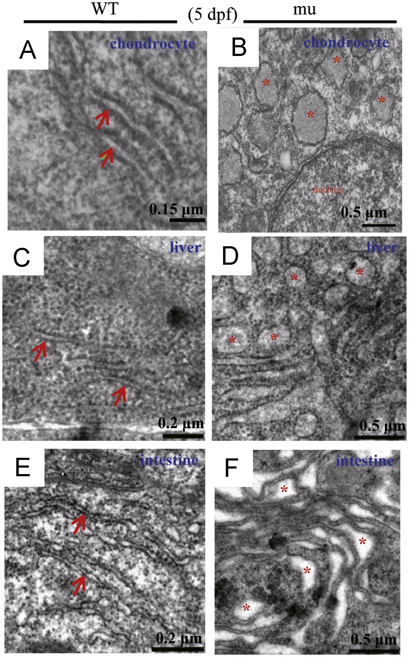
Figure Caption
Fig. 5 The sq198 mutation disrupts the ER structure in chondrocytes, hepatocytes and intestinal epithelium. (A–F) TEM analysis of the ER structure in chondrocytes (A,B), hepatocytes (C,D), and intestinal epithelium (E,F) in wildtype (WT) and mutant (mu) embryos at 5 dpf. Rough ER in WT cells are indicated with red arrows (A,C,E), disrupted ER structures in the mutant cells are highlighted with red asterisks (B,D,F).
Figure Data
Acknowledgments
This image is the copyrighted work of the attributed author or publisher, and
ZFIN has permission only to display this image to its users.
Additional permissions should be obtained from the applicable author or publisher of the image.
Reprinted from Developmental Biology, 367(2), Niu, X., Gao, C., Jan Lo, L., Luo, Y., Meng, C., Hong, J., Hong, W., and Peng, J., Sec13 safeguards the integrity of the endoplasmic reticulum and organogenesis of the digestive system in zebrafish, 197-207, Copyright (2012) with permission from Elsevier. Full text @ Dev. Biol.