
Image
|
Figure Caption
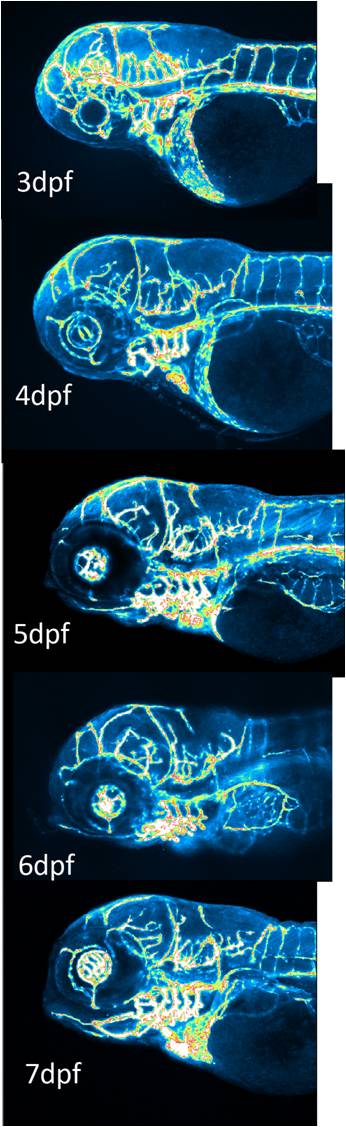
Fig. S1 Comparison of cerebrovascular structures in control zebrafish embryos at 3–7 dpf. Embryos expressed GFP in endothelial cells and images were captured with a confocal microscope.
Figure Data
Acknowledgments
This image is the copyrighted work of the attributed author or publisher, and
ZFIN has permission only to display this image to its users.
Additional permissions should be obtained from the applicable author or publisher of the image.
Full text @ PLoS One