
Image
|
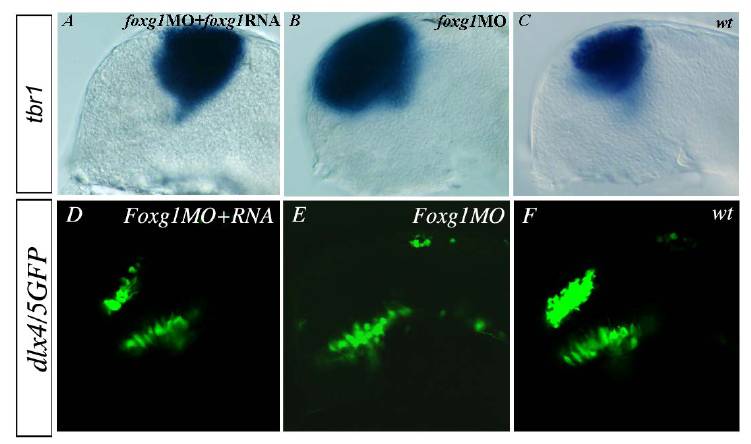
Figure Caption
Fig. S8
foxg1 RNA Injection Rescues foxg1 Morphant Phenotype
All lateral views of prim-5 embryos. The tbr1 expansion normally observed in the foxg1MO (B) is rescued by co-injection with Xenopus foxg1RNA (A, n=17/17). Subpallial neuronal cell types are also rescued as visualised by the presence of GFP positive cells (D, n = 21/21) in Tg(dlx4/5:GFP) embryos (D), while these cells are lacking in single foxg1MO (E), showing that foxg1 morphant phenotype is strictly due to loss of Foxg1 proteins.
Acknowledgments
This image is the copyrighted work of the attributed author or publisher, and
ZFIN has permission only to display this image to its users.
Additional permissions should be obtained from the applicable author or publisher of the image.
Reprinted from Developmental Cell, 16(4), Danesin, C., Peres, J.N., Johansson, M., Snowden, V., Cording, A., Papalopulu, N., and Houart, C., Integration of telencephalic Wnt and hedgehog signaling center activities by Foxg1, 576-587, Copyright (2009) with permission from Elsevier. Full text @ Dev. Cell