
|
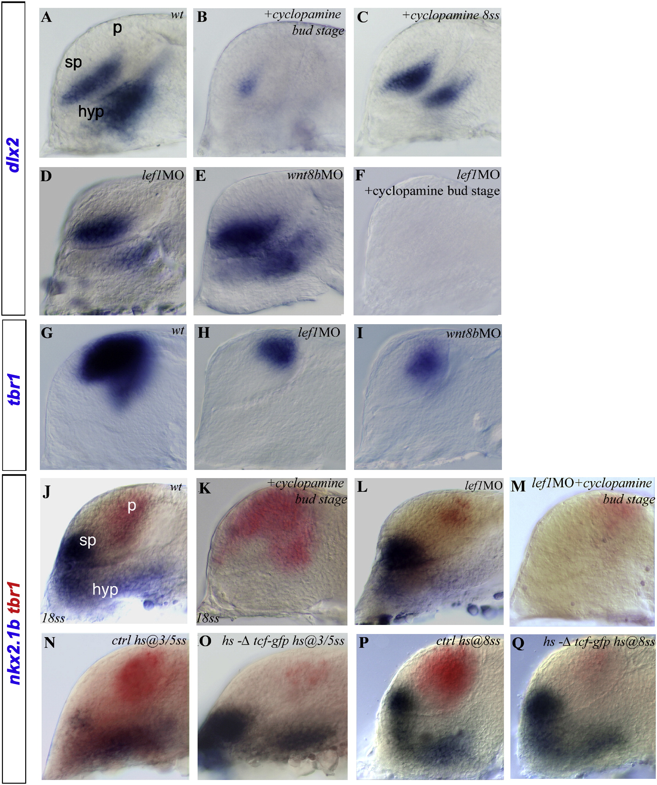
Fig. 1 Shh and Wnt Signaling Activities Independently Regulate the Establishment of the Dorsoventral Axis in the Telencephalon(A–Q) Here, as in all subsequent panels, embryos are shown in lateral view, with anterior oriented toward the left and dorsal oriented toward the top unless stated otherwise. Treatments and/or MO injected in embryos are indicated in the top right corners. Embryos are at the 22ss, except when stated otherwise (bottom left of the picture). Expression of (A–F) dlx2 and (J–Q) nkx2.1b (blue) and of (G–I and J–Q) tbr1 (red and blue) in (A, G, J, N, and P) WT/control, cyclopamine-treated embryos at the (B and K) bud or the (C) 8ss, (D, H, and L) the lef1 morphant, the (E and I) wnt8b morphant, (F and M) lef1 morphants treated with cyclopamine at the bud stage, and hs-Δtcf-gfp transgenic embryos heat shocked at the (O) 3ss and the (Q) 8ss embryos. hyp, hypothalamus; sp, subpallium; p, pallium.
Reprinted from Developmental Cell, 16(4), Danesin, C., Peres, J.N., Johansson, M., Snowden, V., Cording, A., Papalopulu, N., and Houart, C., Integration of telencephalic Wnt and hedgehog signaling center activities by Foxg1, 576-587, Copyright (2009) with permission from Elsevier. Full text @ Dev. Cell