
|
Fig. 5
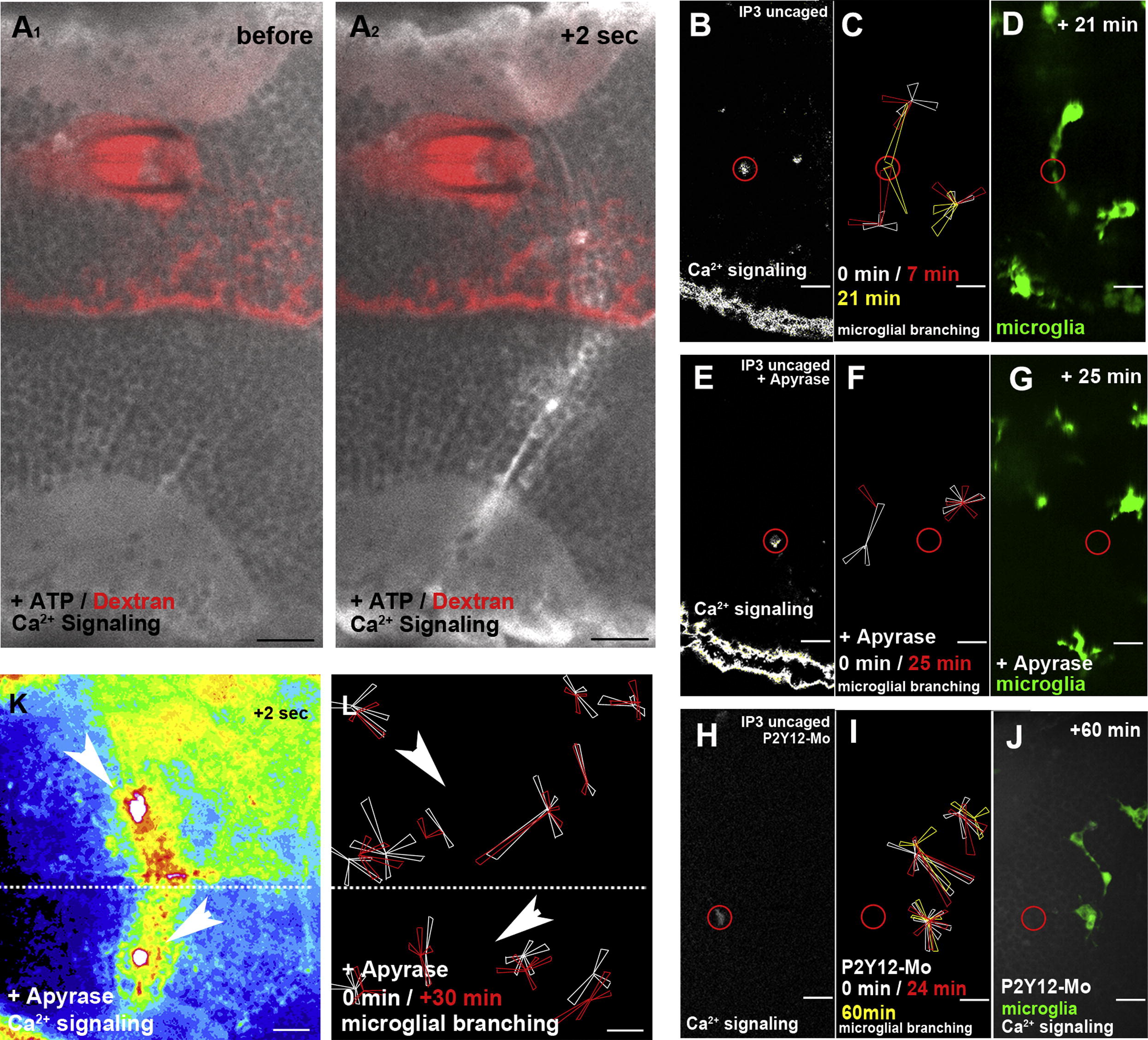
ATP Does Not Influence Ca2+ Wave Formation but Promotes Microglial Movement(A1) Injection of ATP does not promote Ca2+ wave formation (beta-actin::GCaMP3.1).(A2) Same ATP-injected embryo as in (A1) shows normal Ca2+ wave formation upon laser injury.(B) Ca2+ signaling (beta-actin::GCAMP3.1) after IP3 uncaging (Movie S6, left). The location of the uncaging is marked with a red circle.(C) Rose plot of microglial response to IP3 uncaging showing images before uncaging (white) and 7 min (red) and 21 min (yellow) after uncaging.(D) Microglial response (pU1::Gal4-UAS-TagRFP) in the same embryo as in (C) 21 min after IP3 uncaging (Movie S6, right).(E) Ca2+ signaling (beta-actin::GCAMP3.1) upon IP3 uncaging in an embryo additionally injected with apyrase (Movie S7, upper left).(F) Rose plot of microglial response to IP3 uncaging in an apyrase-injected brain showing images before (white) and 25 min after (red) uncaging.(G) Microglial response (pU1::Gal4-UAS-TagRFP) in the same embryo as in (F) 25 min after IP3 uncaging (Movie S7, upper right).(H) Ca2+ signaling (beta-actin::GCAMP3.1) upon IP3 uncaging in an embryo injected with P2Y12-Mo (Movie S7>, lower left).(I) Rose plot of microglial response to IP3 uncaging in a P2Y12-depleted brain showing microglia positions before (white) and 24 min (red) and 60 min (yellow) after uncaging.(J) Microglial response (pU1::Gal4-UAS-TagRFP) in the same embryo as in (I) 60 min after IP3 uncaging (
Reprinted from Developmental Cell, 22(6), Sieger, D., Moritz, C., Ziegenhals, T., Prykhozhij, S., and Peri, F., Long-Range Ca(2+) Waves Transmit Brain-Damage Signals to Microglia, 1138-1148, Copyright (2012) with permission from Elsevier. Full text @ Dev. Cell