
|
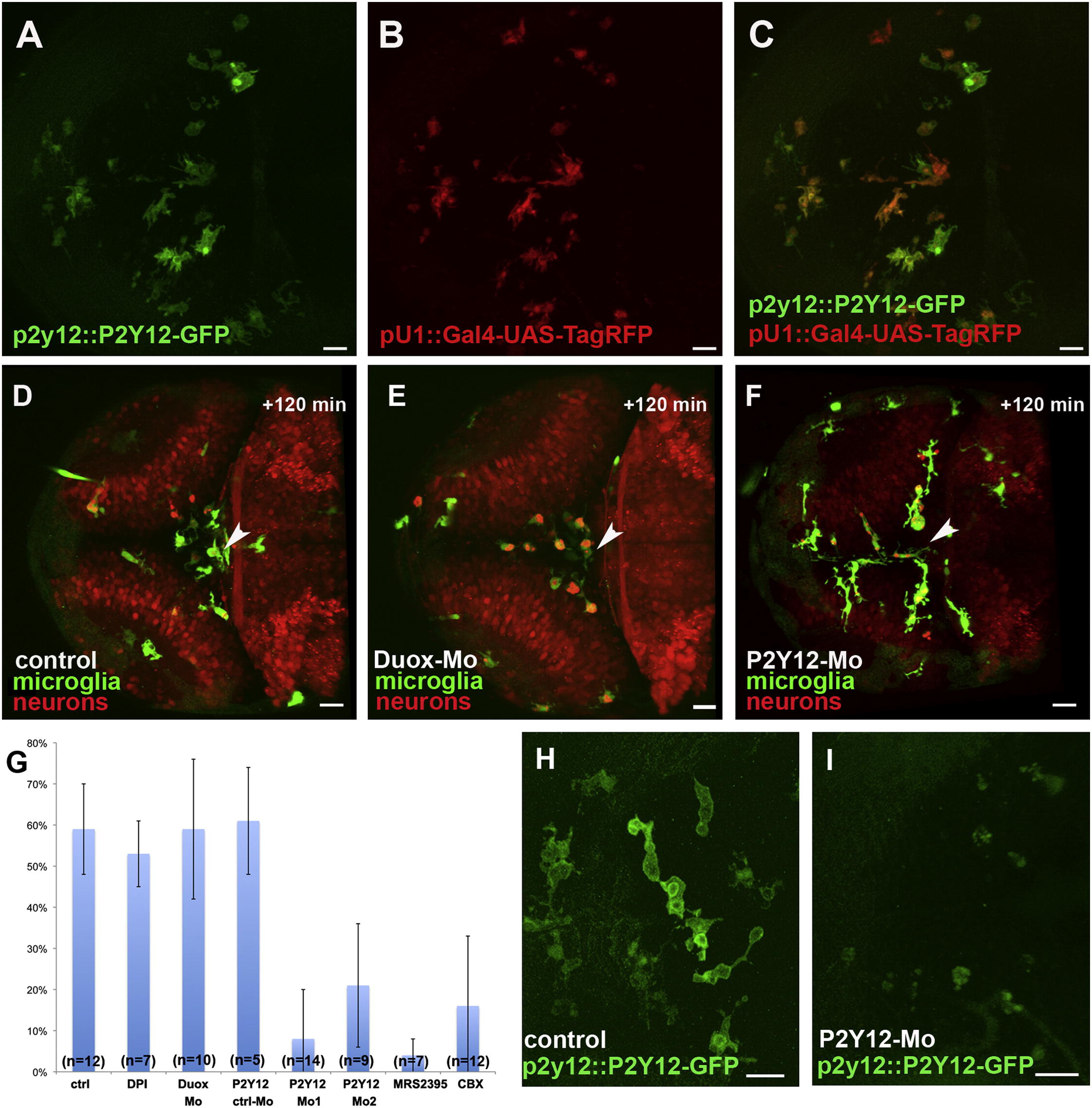
Fig. 4 Microglial Movement Is Controlled via P2Y12 Activation(A) Microglial cells express a P2Y12-GFP fusion protein under the endogenous P2Y12 promoter (p2y12::P2Y12-GFP).(C) Overlay of (A) and (B).(D–F) microglia (pU1::Gal4-UAS-GFP) and neurons (NBT::DsRed) in wild-type (D), Duox-morpholino-injected (E), and P2Y12-morpholino-injected embryos (F) 120 min after central injury. The sites of injury are marked with white arrowheads.(G) Percentage of microglia in the optic tectum moving to the injury site in control (n = 12), DPI-treated (n = 7), Duox morphant (n = 10), MRS2395-treated (n = 7), CBX-treated (n = 12), P2Y12-Mo1-injected (n = 14), P2Y12 Mo2-injected (n = 9), and P2Y12 ctrl Mo-injected (n = 5) brains. Error bars represent standard deviations.(H and I) Dorsal views of a 3 dpf larval brain, showing microglia in the p2y12::P2Y12-GFP line (H) and how P2Y12-morpholino injection strongly decreases P2Y12-GFP expression in these microglia (I).Scale bars for all images: 20 μm. All images were produced using an Olympus FV 1000 with a 40×/NA1.15 objective.See also Figure S2 and Movies S4 and S5.
Reprinted from Developmental Cell, 22(6), Sieger, D., Moritz, C., Ziegenhals, T., Prykhozhij, S., and Peri, F., Long-Range Ca(2+) Waves Transmit Brain-Damage Signals to Microglia, 1138-1148, Copyright (2012) with permission from Elsevier. Full text @ Dev. Cell