
|
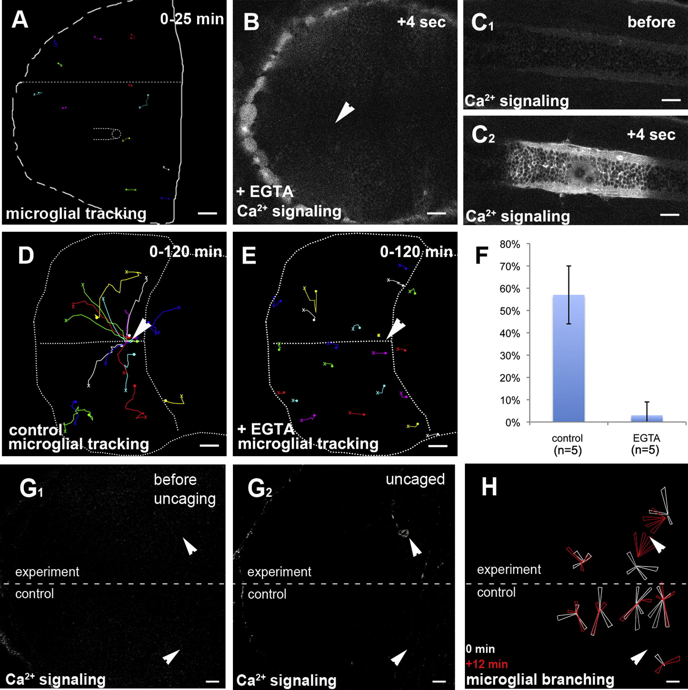
Fig. 3 Ca2+ Signaling Prefigures and Guides Microglial Migration(A) Microglial tracking (pU1::Gal4-UAS-TagRFP) upon control injection of water in a 3 dpf larval brain. The starting point of individual tracks is marked with an X and the end point with a dot.(B) Injection of EGTA blocks the formation of Ca2+ waves (beta-actin::GCAMP3.1) upon laser injury (Movie S3, middle). The site of injury is marked with a white arrowhead.(C1 and C2) Control injury in the trunk of the same embryo as in (B). Ca2+ imaging (beta-actin::GCAMP3.1) before (C1) and after (C2) injury (Movie S3, right).(D) Cell tracking of microglia (pU1::Gal4-UAS-TagRFP) upon central brain injury in a control embryo. The white arrowhead marks the site of injury. The starting point of individual tracks is marked with an X and the end point with a dot.(E) Cell tracking of microglia (pU1::Gal4-UAS-TagRFP) upon central brain injury in a EGTA injected embryo. The site of injury is marked with a white arrowhead. The starting point of individual tracks is marked with an X and the end point with a dot.(F) Quantification of microglia movement to an injury site (percentage of microglia in the optic tectum responding to injury) in control (left, n = 5) and EGTA-injected brains (right, n = 5). Error bars represent standard deviations.(G) Single-hemisphere injections of caged IP3 (experiment). Single-neuron uncaging leads to Ca2+ signaling (beta-actin::GCaMP3.1) in the injected, but not in the control, side. Laser uncaging sites before (G1) and after (G2) uncaging are marked by white arrowheads.(H) Rose plot of microglial response to IP3 uncaging, showing microglia positions before (white) and 12 min after (red) uncaging.Scale bars for all images: 20 μm. Images in (B)–(F) were produced using an Olympus FV 1000 with a 40×/NA1.15 objective. Images in (A) and (G)–(H) were done using an Andor Spinning Disk Confocal with a 20×/NA0.7 objective.See also Figure S1 and Movies S3, S4, and S6.
Reprinted from Developmental Cell, 22(6), Sieger, D., Moritz, C., Ziegenhals, T., Prykhozhij, S., and Peri, F., Long-Range Ca(2+) Waves Transmit Brain-Damage Signals to Microglia, 1138-1148, Copyright (2012) with permission from Elsevier. Full text @ Dev. Cell