
|
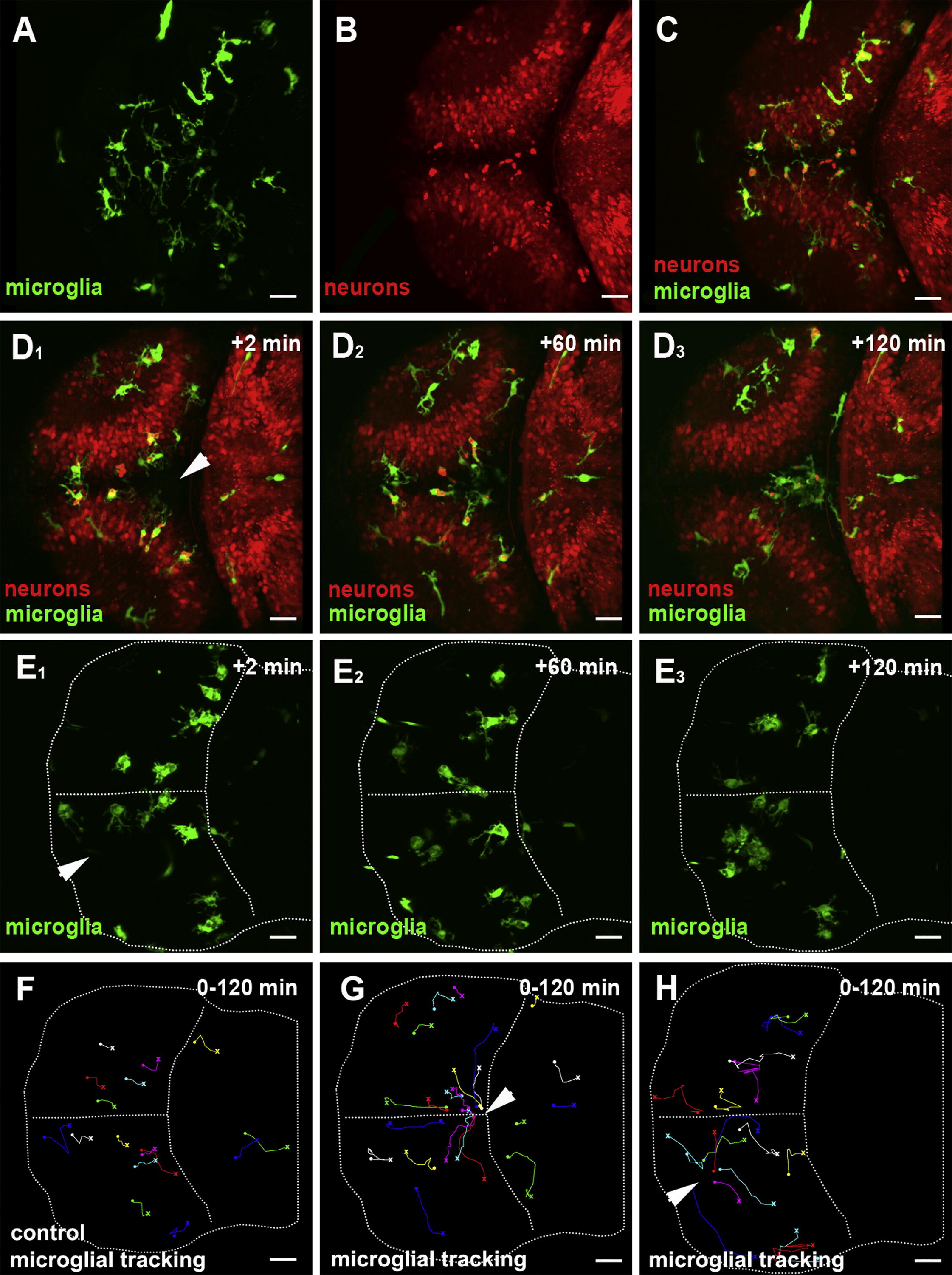
Fig. 1 Microglial Migration upon Laser-Induced Neuronal Injuries(A) Dorsal view of a 3 dpf larval brain. Microglia (pU1::Gal4-UAS-GFP).(B) Dorsal view of a 3 dpf larval brain. Neurons (NBT::DsRed).(C) Overlay of (A) and (B).(D1–D3) Confocal time-lapse showing microglia (green) responding to a central injury (recording times indicated) (Movie S1, left). The injury site is marked with a white arrowhead.(E1–E3) Confocal time-lapse showing microglia (green) responding to a single-hemisphere injury (recording times indicated) (Movie S1, right). The injury site is marked with a white arrowhead.(F) Microglial cell tracking over 120 min in the preinjured brain. The starting point of individual tracks is marked with an X and the end point with a dot.(G) Microglial cell tracking over 120 min upon central injury. The injury site is marked with a white arrowhead.(H) Microglial cell tracking over 120 min upon single-hemisphere injury. The injury site is marked with a white arrowhead.Scale bars for all images: 20 μm. All images were produced using an Olympus FV 1000 with a 40×/NA 1.15 objective.See also Figure S1and Movie S1.
Reprinted from Developmental Cell, 22(6), Sieger, D., Moritz, C., Ziegenhals, T., Prykhozhij, S., and Peri, F., Long-Range Ca(2+) Waves Transmit Brain-Damage Signals to Microglia, 1138-1148, Copyright (2012) with permission from Elsevier. Full text @ Dev. Cell