
Image
|
Figure Caption
Fig. S1
Neuronal injuries and the formation of Ca2+ waves, related to Figure 3
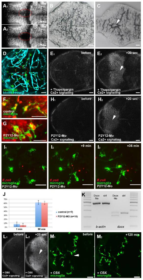
(A1 and A2) Dorsal views of a 3 dpf larval brain, showing neurons (NBT::DsRed) before (A1) and after (A2) injury. (B and C) Histological sections of an un-injured (B) and injured brain (C). The lesion is marked with a white arrowhead. (D) Dorsal view of a 3 dpf larval brain showing microglia in green (pU1::Gal4-UAS-TagRFP) and the vasculature in blue (Fli::eGFP). (E1 and E2) Time lapse of Ca2+ signaling (beta-actin::GCaMP3.1) in a Thapsigargin injected brain before (E1) and after (E2) laser ablation (white arrowhead). (F) Microglia (pU1::Gal4-UAS-GFP) engulf neurons (NBT::DsRed) as shown by the red neuronal material present within the cell. (G) P2Y12 morphant microglia engulf red neuronal material as control cells. (H1 and H2) Time lapse of Ca2+ signaling (beta-actin::GCaMP3.1) in a P2Y12-Mo injected embryo before (H1) and after (H2) laser ablation (white arrowhead). (I1-I3) Time lapse of the microglial response (pU1::Gal4-UAS-GFP) towards bacteria (E. coli) in a P2Y12-Mo injected embryo (Mov.S5). (J) Percentage of microglia in a 100 μm wide circle around the side of injection having engulfed bacteria in control and P2Y12 injected embryos 1 min and 90 min post infection. Error bars represent standard deviations. (K) RT-PCR showing the splice effect of the Duox-Mo. (L1 and L2) Time lapse of Ca2+ signaling (beta-actin::GCaMP3.1) in a CBX injected brain before (L1) and after (L2) laser ablation (white arrowhead). (M1 and M2) microglia (pU1::Gal4-UAS-GFP) before (M1) and 120 min after (M2) central brain injury (white arrowhead) in CBX injected brains. Scale bars always: 20 μm. Images (A, D-H, L, M) were done using an Olympus FV 1000 with a 40X/NA1.15 objective. Images (I1-I3) were done using an Andor Spinning Disk Confocal with a 20x/NA0.7 objective. Images (B, C) were done using a Zeiss Axiophot with a 20X/NA0.5 objective.
Acknowledgments
This image is the copyrighted work of the attributed author or publisher, and
ZFIN has permission only to display this image to its users.
Additional permissions should be obtained from the applicable author or publisher of the image.
Reprinted from Developmental Cell, 22(6), Sieger, D., Moritz, C., Ziegenhals, T., Prykhozhij, S., and Peri, F., Long-Range Ca(2+) Waves Transmit Brain-Damage Signals to Microglia, 1138-1148, Copyright (2012) with permission from Elsevier. Full text @ Dev. Cell