
|
Fig. 7
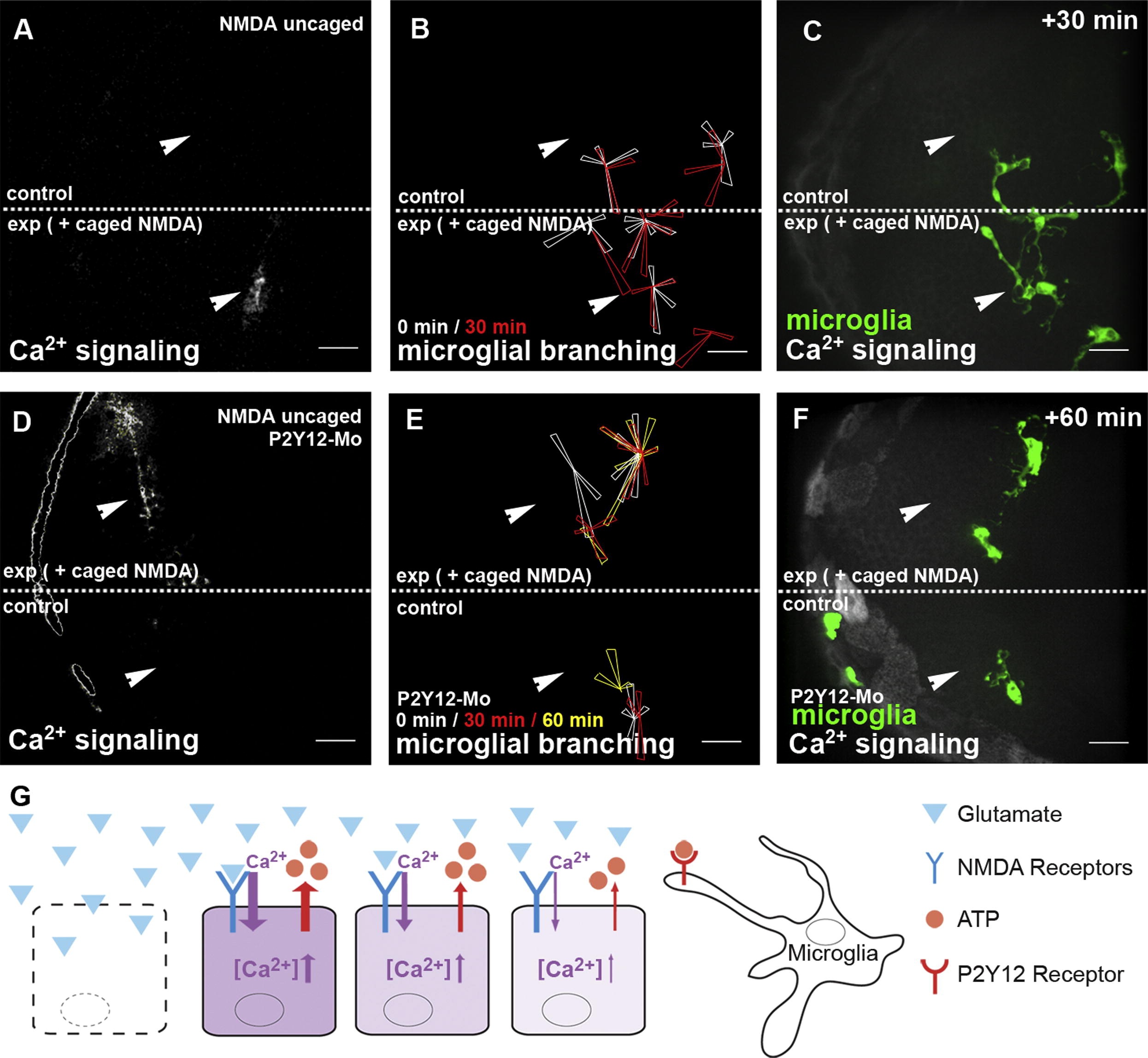
Microglial Branching toward Sites of Glutamate Signaling Is Mediated via the Microglial ATP Receptor P2Y12(A) Single-hemisphere injections of caged NMDA. Uncaging leads to Ca2+ signaling (beta-actin::GCaMP3.1) in the injected, but not in the control, side (Movie S11, upper). Laser uncaging sites are marked by white arrowheads.(B) Rose plot of microglial response to NMDA uncaging, showing microglia positions before (white) and 30 min after (red) uncaging.(C) Microglial response (pU1::Gal4-UAS-TagRFP) in the same embryo as in (B) 30 min after NMDA uncaging (Movie S11, upper).(D) Single-hemisphere injections of caged NMDA in P2Y12-Mo-injected embryos. Laser uncaging sites are marked by white arrowheads.(E) Rose plot of microglial response to NMDA uncaging in P2Y12-depleted brains showing microglia positions before (white) and 30 min (red) and 60 min (yellow) after uncaging.(F) Microglial response (pU1::Gal4-UAS-TagRFP) in the same embryo as in (E) 60 min after NMDA uncaging (
Reprinted from Developmental Cell, 22(6), Sieger, D., Moritz, C., Ziegenhals, T., Prykhozhij, S., and Peri, F., Long-Range Ca(2+) Waves Transmit Brain-Damage Signals to Microglia, 1138-1148, Copyright (2012) with permission from Elsevier. Full text @ Dev. Cell