
|
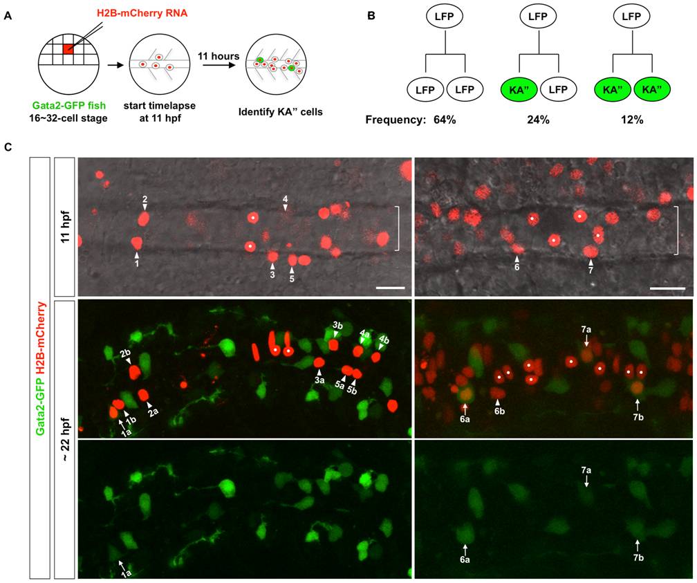
Fig. 2 Lineage analysis of the LFP domain.
(A) Schematic drawings of scatter labeling and time-lapse imaging. Gata2-GFP fish were injected with H2B-mCherry mRNA (red) into a single blastomere at 16- to 32-cell stage. Scatter labeled embryos with nuclear mCherry expression (red) were imaged in the dorsal view starting at 3-somite stage (11 hpf) for about 11 hours. At the end of the time-lapse, an image with both the green and red channels was acquired to identify Gata2-GFP-expressing KA′′ cells (green). (B) Observed division patterns in the LFP domain. Of a total of 25 cell divisions tracked, 16 were LFP/LFP divisions, 6 were KA′′/LFP divisions, and 3 were KA′′/KA′′ divisions. KA′′ cells (green) were identified base on the expression of Gata2-GFP reporter. (C) Gata2-GFP fish (green) was scatter labeled by H2B-mCherry (red) and imaged from 11 hpf for about 11 hours. Two examples are shown. Top panel: merged images with both the red channel and the bright field of a single optical slice at the start of the movie at 11 hpf. The underlying notochord (brackets) is visible but out of focus. Middle and bottom panels: the merged image with both green and red channels and the green channel alone of a confocal projection at the end of the movie around 22 hpf. KA′′ cells (arrows) can be distinguished from LFP cells (arrowheads) based on the expression of Gata2-GFP (green) at 22 hpf. Medial floor plate cells are indicated by white dots. Lineage related cells confirmed by cell tracking are indicated (also see Figure S1 and Video S1). For example, cell 1 generates a KA′′ cell (cell 1a) and an LFP cell (cell 1b). Of the 7 examples shown here, cells 1 and 6 undergo KA′′/LFP divisions, cell 7 undergoes KA′′/KA′′ division, and cells 2–5 divide symmetrically giving rise to two LFP cells (LFP/LFP divisions). Note that cells 4 and 6 are more dorsally located and therefore not in focus in images at 11 hpf (top panels). Scale bars: 20 μm.