
|
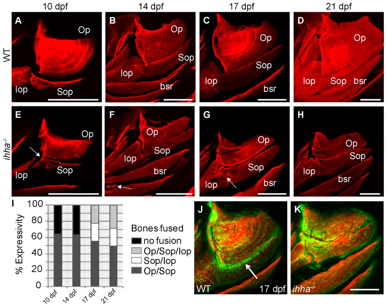
Fig. 9 ihha is required for proper joint formation between dermal bones. Images are lateral views with dorsal upwards and anterior towards the left. (A-H) Confocal projections of live wild-type and ihha mutant larvae stained with Alizarin Red. (A-D) Wild-type Op and adjacent bones from 10 to 21 dpf. (E) ihha mutant at 10 dpf with reduced ventral length and fusions between the ventral Op edge and dorsal Sop edge (arrow). (F) ihha mutant at 14 dpf with a nearly complete fusion of the Op and Sop, and ectopic ossification between the branchiostegal rays (arrow). (G) ihha mutant at 17 dpf with an Sop-Iop fusion (arrow), but no Op-Sop fusion. (H) ihha mutant at 21 dpf showing Iop-Sop fusion and reduced Op size. (I) Representation of the percentage expressivity of each fusion phenotype apparent in a single clutch of ihha mutants from 10 to 21 dpf. (J,K) Confocal projections of the Op stained with Alizarin Red in larvae expressing the trps1:EGFP transgene (green) as a joint marker. (J) High levels of trps1:EGFP expression are detectable in the dorsal Op joint and immediately ventral to the vp edge (arrow), overlapping the Sop. (K) ihha mutants with Op-Sop fusions show decreased trps1:EGFP expression along the presumed vp edge where the bones are now fused. bsr, branchiostegal ray; Iop, interopercle; Op, opercle; Sop, subopercle. Scale bars: 200 μm.