
|
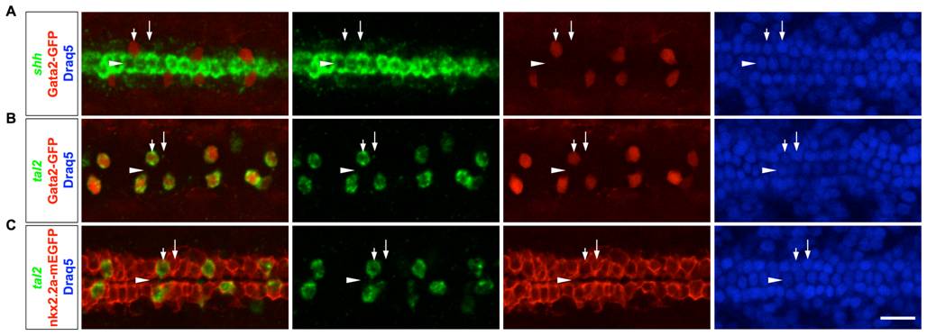
Fig. S2 LFP cells can be reliably identified based on their locations. (A) Gata2-GFP embryos were stained with the shh probe (green), the GFP antibody (red) and the Draq5 dye (blue) to label cell nuclei. Gata2-GFP-positive KA′′ cells (short arrows) and Gata2-GFP-negative LFP cells (long arrows) flank the shh-expressing medial floor plate cells (arrowheads). (B) Gata2-GFP embryos were stained with the tal2 probe (green), the GFP antibody (red) and the Draq5 dye (blue). All Gata2-GFP-positive KA′′ cells (short arrows) also express tal2. (C) nkx2.2a-mEGFP embryos were stained with the tal2 probe (green), the GFP antibody (red) and the Draq5 dye (blue). All tal2-negative cells (long arrows) immediately flanking the medial floor plate (arrowheads) are LFP cells, indicated by the expression of membrane localized EGFP under the control of the nkx2.2a promoter (nkx2.2a-mEGFP). Dorsal views of embryos at 21 hpf are shown. Scale bars: 20 μm.