
|
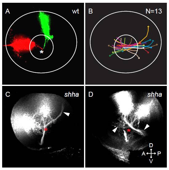
Fig. S1 Focal dye injections show prevalence for posterior RGC axon projections in shha mutants. Maximum-intensity confocal projections of lateral views of focal lipophilic dye injections into the retina of 2 dpf wt and shha embryos. (A) Wt eye injected with DiI anterior and DiO dorsal shows axon projections towards the optic disc (white asterisk), where the axons leave the eye through the optic nerve. (B) Diagram showing shha RGC axon projections as seen after DiI injections of 13 embryos. Axon bundles from all quadrants of the retina project towards the optic disc but fail to leave through the optic nerve and instead continue growing inside the eye. Posterior RGC projections are seen more commonly than anterior projections. (C,D) Examples of shha eyes focally injected with DiI. Misprojecting RGC axon bundles grow towards the optic disc (red asterisk) before projecting posteriorly or anteriorly (arrowheads). D, dorsal; V, ventral; A, anterior; P, posterior.