
|
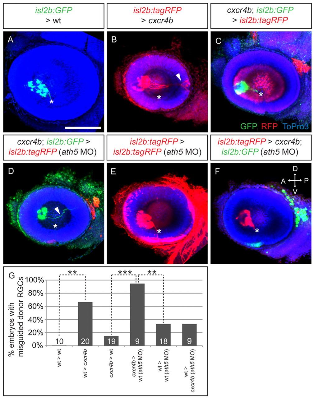
Fig. 8 Cxcr4b acts cell autonomously in RGC for correct intraretinal axon pathfinding. (A-F) Maximum-intensity projections of lateral views of 54 hpf zebrafish embryos transplanted with donor cells at 24 hpf. (A) Wt RGC axons (isl2b:GFP) in wt embryos exit the eye (asterisk). (B) Wt RGC axons (isl2b:tagRFP) often make errors in cxcr4b hosts (arrowhead). (C) cxcr4b RGC axons (isl2b:GFP) exit the eye in most cases when transplanted into wt hosts (isl2b:tagRFP). (D) Cxcr4b RGC axons (isl2b:GFP) are misguided in ath5 morphants (isl2b:tagRFP) in almost all transplants (arrowhead). (E,F) Wt RGC axons (isl2b:tagRFP) rarely make errors in ath5 morphants (isl2b:tagRFP) (E)and in cxcr4b ath5 morphants (isl2b:GFP) (F). D, dorsal; V, ventral; A, anterior; P, posterior. Scale bar: 100 μm. (G) Percentage of host embryos with misguided donor RGC axons. Number of embryos shown at base of bars. **P<0.01, ***P<0.001, Fisher′s exact test.