
|
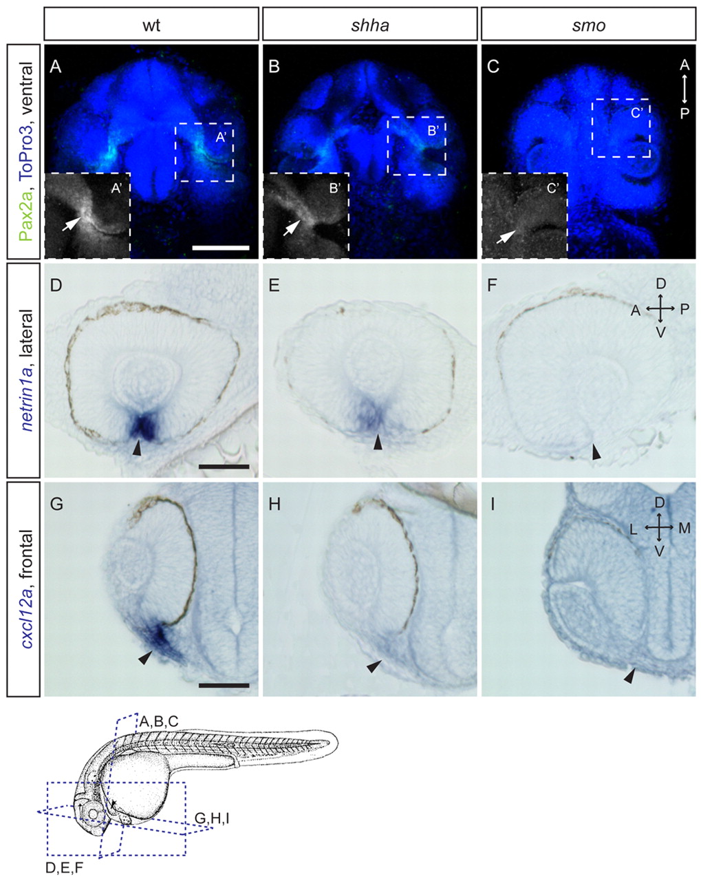
Fig. 6 Expression of optic stalk markers is decreased in Hh mutants. (A-C2) Maximum-intensity projections of zebrafish embryos stained for Pax2a (green) by immunohistochemistry (28 hpf); nuclei, ToPro3 (blue). Ventral views. Pax2a reduced in shha (B) and absent in smo (C) in optic stalk (arrow) compared with wt (A). Insets (A2-C2) show magnified optic stalk region, Pax2a staining only. (D-F) Whole-mount in situ hybridizations (15 μm sagittal sections; 28 hpf). netrin1a at the optic fissure (arrowheads) is decreased in shha (E) and lost in smo (F) compared with wt (D). (G-I) Coronal sections of whole mount in situ hybridizations (28 hpf). cxcl12a expression in optic stalk (arrowheads) is reduced in shha (H) and lost in smo (I) compared with wt (G). D, dorsal; V, ventral; A, anterior; P, posterior. Illustration below shows plane of views for panels above. Adapted with permission (Kimmel et al., 1995). Scale bars: 100 μm.