
|
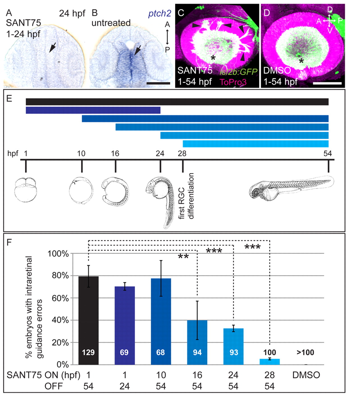
Fig. 5 Shh is required during eye patterning for intraretinal axon pathfinding in zebrafish. SANT75 (40 μM) was bath applied to inhibit Hh signaling for specific stages of embryonic development. (A,B) Whole-mount in situ hybridizations (15 μm sections), dorsal views. ptch2 mRNA is expressed at the midline (arrow) in DMSO-treated embryos (24 hpf) (B), whereas expression is lost (arrow) after SANT75 treatment (1-24 hpf) (A). (C,D) Maximum-intensity projections of lateral views (54 hpf). DMSO (1%) (D) does not affect RGC axon projections in isl2b:GFP embryos (green), whereas 40 μM SANT75 (1-54 hpf) (C) yields misguided RGC axons (arrowheads). Nuclei, ToPro3 (magenta); optic disc indicated by asterisk. (E,F) Different SANT75 application time points (E) and percentage of embryos with resulting intraretinal guidance errors (F). Number of embryos shown at base of bars. Error bars represent s.d. ne3 experiments, **P<0.01, ***P<0.001, Student′s t-test. Line drawings (E) adapted with permission (Kimmel et al., 1995). D, dorsal; V, ventral; A, anterior; P, posterior. Scale bars: 100 μm.