
|
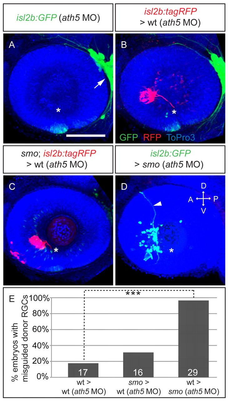
Fig. 4 Transplants into RGC-free hosts confirm non-cell-autonomous effect of Smo in intraretinal axon pathfinding. (A-D) Maximum-intensity projections of 54 hpf isl2b:GFP or isl2b:tagRFP zebrafish embryos injected with ath5 MO at 1-cell stage (A) and transplanted with donor cells at 24 hpf (B-D). (A) No RGC differentiation in ath5 morphants; trigeminal ganglion as control for transgene expression (arrow). (B) Wt RGC axons (isl2b:tagRFP, red) in isl2b:GFP (green) ath5 morphants rarely make errors. (C) smo (isl2b:tagRFP) RGC axons in isl2b:GFP ath5 morphants are rarely misguided. (D) Wt (isl2b:GFP) RGC axons in smo ath5 morphants make errors (arrowhead). Asterisk indicates optic disc. Nuclei, ToPro3 (blue). D, dorsal; V, ventral; A, anterior; P, posterior. Scale bar: 100 μm. (E) Percentage of embryos with misrouted axons. Numbers of embryos shown at base of bars. ***P<0.005, Fisher′s exact test.