
|
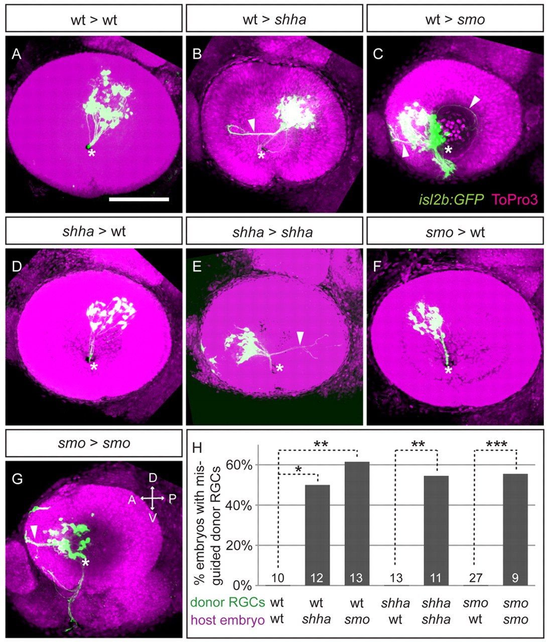
Fig. 3 Shh and Smo act non-cell-autonomously in intraretinal axon pathfinding in zebrafish. (A-G) Representative images of host eyes at 54 hpf after cell transplants at 24 hpf. Lateral views of maximum-intensity projections. Wt RGCs axons exit the eye through the optic disc (asterisk) in wt hosts (A), but often misproject (arrowheads) in shha (B) and smo (C) hosts. Shha RGC axons always exit the eye in wt hosts (D), whereas many misproject in shha hosts (E). Similar results found with smo RGCs in wt (F) or smo (G) hosts. Transplanted RGCs are isl2b:GFP (green) or isl2b:tagRFP (pseudocolored green in F,G); nuclei, ToPro3 (magenta). D, dorsal; V, ventral; A, anterior; P, posterior. Scale bar: 100 μm. (H) Percentage of embryos with misrouted donor RGC axons. Numbers of embryos shown at base of bars. *P<0.05, **P<0.01, ***P<0.001, Fisher′s exact test.