
|
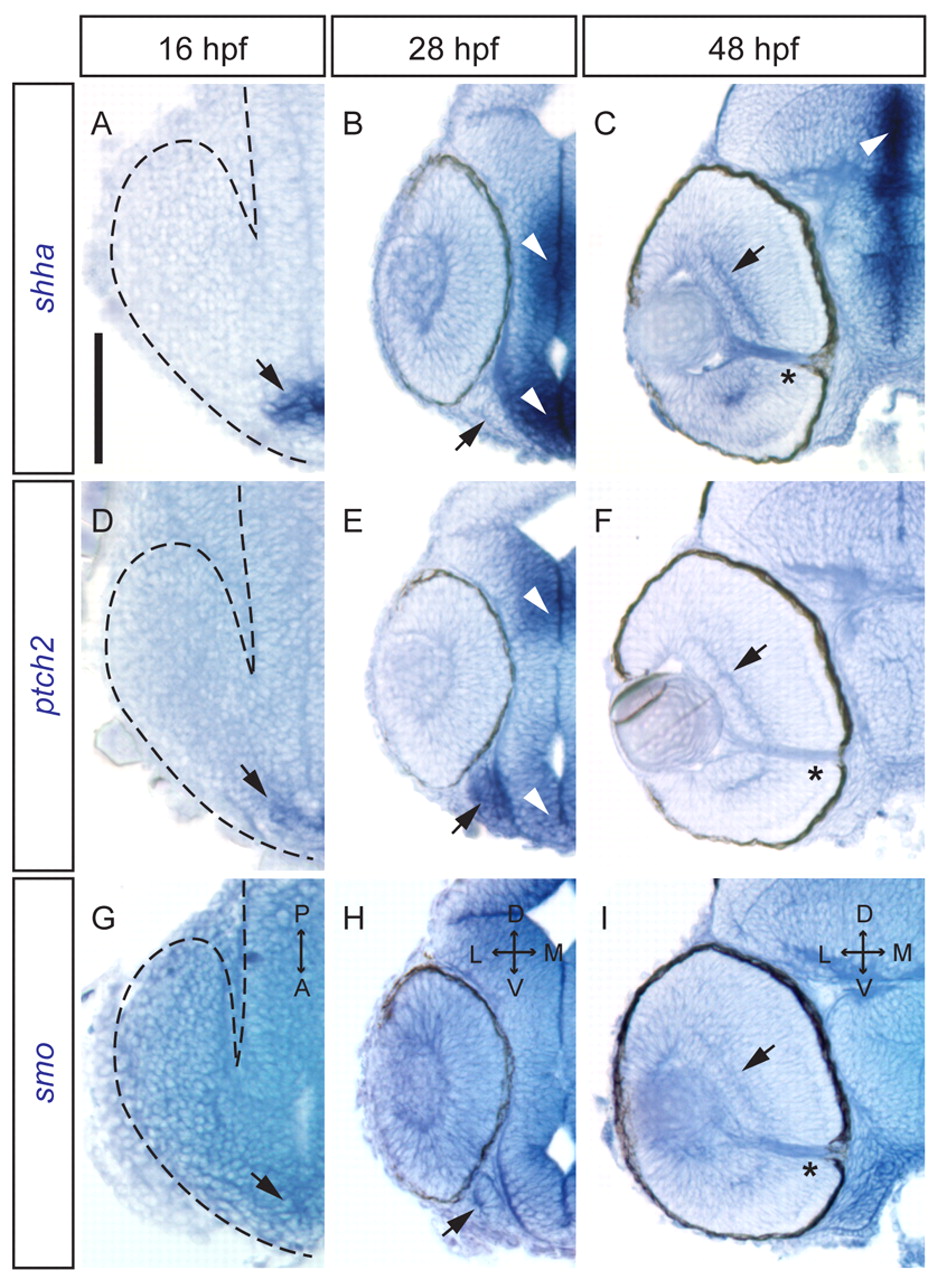
Fig. 2 Hh pathway genes are expressed in the zebrafish optic stalk and RGC layer. Whole-mount in situ hybridizations (15 μm sections) for shha, ptch2 and smo mRNA. A,D,G are dorsal views; B,C,E,F,H,I are frontal views. (A) 16 hpf, shha expression in anterior midline neurectoderm (arrow). (B) 28 hpf, shha expression at the midline (arrowhead) but not optic stalk (arrow). (C) 48 hpf, shha strongly expressed at the midline (arrowhead) and in RGC layer (arrow). mRNA also detected in the optic nerve (asterisk). (D) 16 hpf, ptch2 expressed at the anterior midline (arrow). (E) 28 hpf, ptch2 strongly expressed in the optic stalk (arrow) and midline (arrowheads). (F) 48 hpf, ptch2 detected in the RGC layer (arrow) and optic nerve (asterisk). (G) 16 hpf, smo expressed throughout head region. (H) 28 hpf, smo broadly expressed, including optic stalk (arrow). (I) 48 hpf, smo localized in the RGC layer (arrow) and optic nerve (asterisk). Dashed line in A,D,G outlines the optic vesicle. A, anterior; P, posterior; D, dorsal; V, ventral; L, lateral; M, medial. Scale bar: 100 μm.