
|
Fig. S5
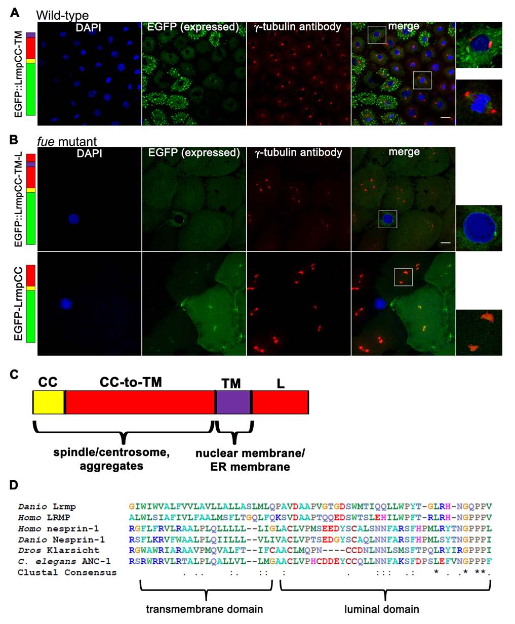
Additional Observations on Fusion Protein Localization and Alignment of the Lrmp C-Terminal Domain to Known KASH Proteins, Related to Figure 5
(A-B) Embryos injected with fusion construct RNAs at the 1-cell stage were fixed at 2.5-3.5 hpf, then labeled with DAPI and anti-γ-tubulin antibody.
(A) Wild-type embryos injected with EGFP::LrmpCC-TM show nuclear membrane and spindle localization of EGFP as well as EGFP aggregation, similar to what is seen with the EGFP::LrmpCC-TM-L fusions.
(B) Localization of overexpressed fusion constructs in fue mutant embryos. Fusion proteins showed subcellular localization patterns similar to those observed when overexpressed in wild-type embryos, namely nuclear membrane localization of the EGFP::LrmpTM-L fusion construct (top row), and centrosomal plus cell membrane localization of the EGFP::LrmpCC-TM construct (bottom row). Scale bars represent 20 μm and apply to all panels in A,B. White boxes indicate regions shown at higher magnification in right-most panels.
(C) Diagram summarizing the roles of the different Lrmp C-terminal subdomains in protein localization.
(D) ClustalW was used to align the C-terminal 60 residues of zebrafish and human LRMP and several KASH proteins from various species. Invariant residues are indicated with an asterisk below the alignment while positions with conserved similarity are marked with a colon (substitutions with highly similar properties) or a period (substitutions with weakly similar properties).