
Image
|
Figure Caption
Fig. 3
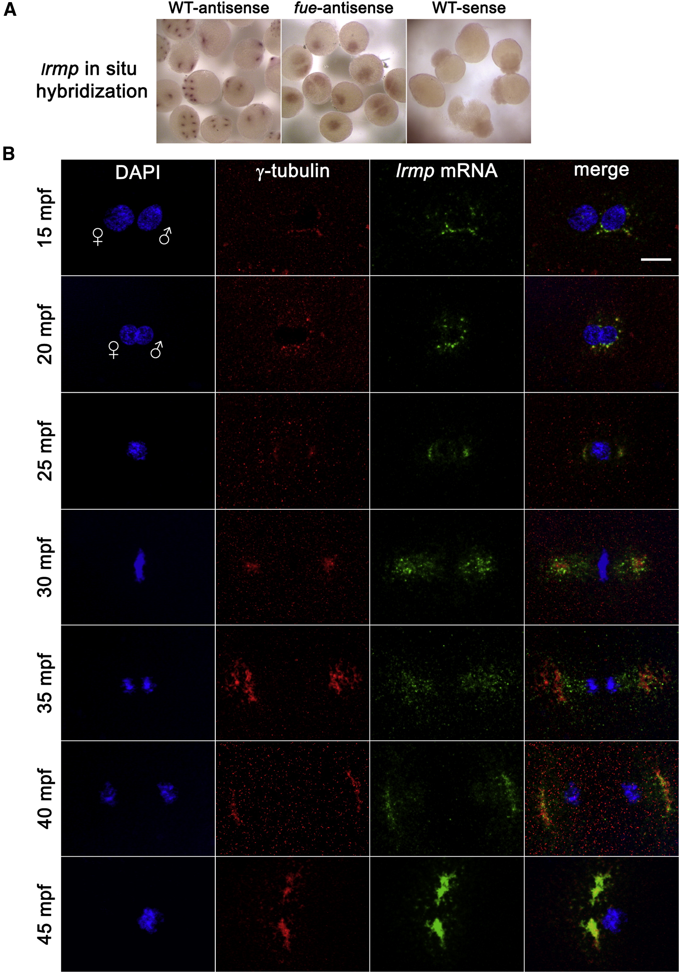
lrmp Transcripts Show Dynamic Localization Patterns Lost in fue Mutants (A) Chromogenic in situ hybridization with lrmp antisense probes in WT (left) and fue mutants (center) and negative control sense probes (right). (B) WT embryos fixed at 5 min intervals and labeled with γ-tubulin antibody (red) and DAPI (blue), in combination with fluorescent in situ detection of lrmp mRNA (green). Scale bar represents 20 μm and applies to all panels in (B). See also Figure S3.
Figure Data
Acknowledgments
This image is the copyrighted work of the attributed author or publisher, and
ZFIN has permission only to display this image to its users.
Additional permissions should be obtained from the applicable author or publisher of the image.
Full text @ Curr. Biol.