
Image
|
Figure Caption
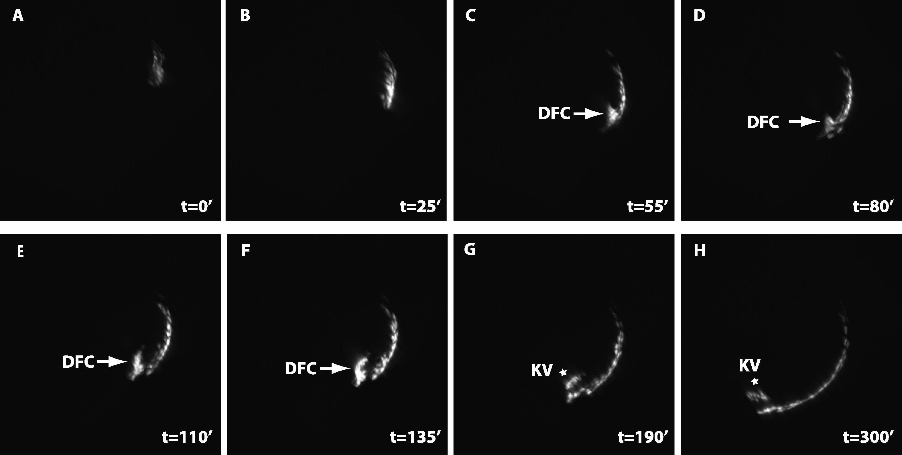
Fig. 2
Dorsal forerunner cells ingress from dorsal EVL to form Kupffer’s vesicles. Time lapse imaging shows that marginal dEVL cells ingress to form dorsal forerunner cells (DFC, arrows), which subsequently form Kupffer’s vesicle (KV, stars). The total duration of the movie is 300 min. The imaging started at the 50% epibody stage (A, t = 02). The interval between each frame is 5 min. Dorsal is to the right.
Figure Data
Acknowledgments
This image is the copyrighted work of the attributed author or publisher, and
ZFIN has permission only to display this image to its users.
Additional permissions should be obtained from the applicable author or publisher of the image.
Reprinted from Mechanisms of Development, 129(1-4), Chen, Y.Y., Harris, M.P., Levesque, M.P., Nüsslein-Volhard, C., and Sonawane, M., Heterogeneity across the dorso-ventral axis in zebrafish EVL is regulated by a novel module consisting of sox, snail1a and max genes, 13-23, Copyright (2012) with permission from Elsevier. Full text @ Mech. Dev.