Fig. 2
Fig. 2
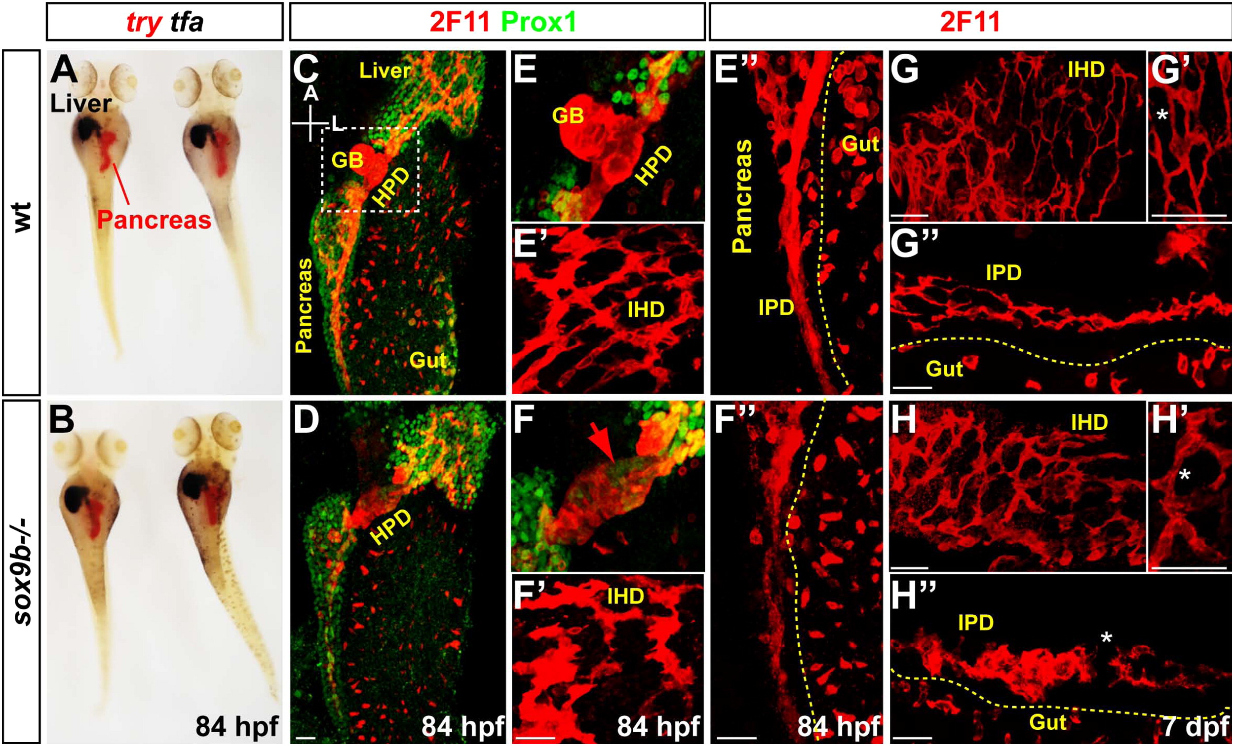
Affected morphogenesis of the IPD, IHD and HPD system in sox9b mutants. (A, B) Acinar (try) and hepatocyte (tfa) differentation as well as the global morphology of the larvae are similar in wild type larvae (A) and in sox9b mutants (B) at 84 hpf. (C, D) Three dimensional rendering of the liver and pancreas at 84 hpf, labelled by Prox1, and of the entire ductal network highlighted by 2F11. The insets represent the 2F11 staining in the liver. White arrows in D and inset point at the disrupted connections between cholangiocytes in sox9b mutants. (E, F) Close-up of the HPD system connecting the pancreas and the liver to the intestine. Ectopic Prox1+ cells are detected throughout the HPD system (red arrow). (E2, F2) Less interconnecting ducts are detected in the IHD sox9b mutants. (E3, F3) 3-D rendering of the pancreas showing weaker 2F11 labelling in the IPD of sox9b mutant (F3) compared to wild type (E3). (G, H). 3-D rendering at 7 dpf of ductal 2F11 labelling in the liver (IHD) in wild type (G) and sox9b mutants (H). (G2, H2) Higher magnification showing thicker connections between IHD cells (asterisk). (G3, H3) 3-D rendering at 7 dpf of ductal 2F11 labelling in the pancreas (IPD) in wild type (G2) and sox9b mutants (H2). GB, gall bladder; HPD, hepatopancreatic ductal system; IHD, intrahepatic ducts; IPD, intrapancreatic ducts. Scale bar=20 μm.
Reprinted from Developmental Biology, 366(2), Manfroid, I., Ghaye, A., Naye, F., Detry, N., Palm, S., Pan, L., Ma, T.P., Huang, W., Rovira, M., Martial, J.A., Parsons, M.J., Moens, C.B., Voz, M.L., and Peers, B., Zebrafish sox9b is crucial for hepatopancreatic duct development and pancreatic endocrine cell regeneration, 268-278, Copyright (2012) with permission from Elsevier. Full text @ Dev. Biol.

