
Fig. 1
- ID
- ZDB-IMAGE-120611-1
- Genes
- Publication
- Chen et al., 2012 - Heterogeneity across the dorso-ventral axis in zebrafish EVL is regulated by a novel module consisting of sox, snail1a and max genes
- All Figures
- Figures for Chen et al., 2012
|
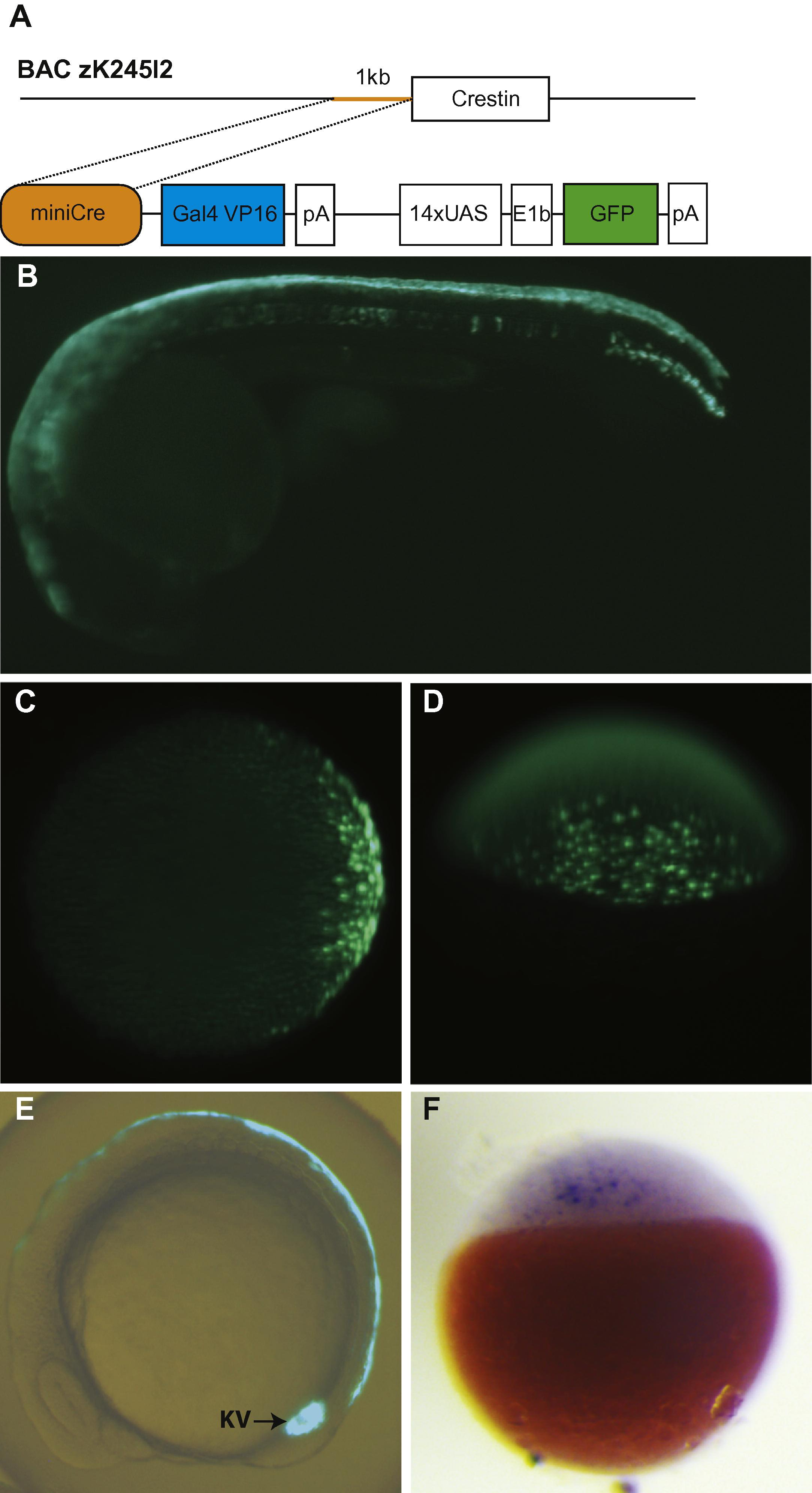
Fig. 1
The miniCrestin promoter drives Gal4-mediated GFP expression in the dorsal EVL and Kupffer’s vesicle. (A) A 1 kb promoter in front of the crestin open reading frame was isolated by PCR from zebrafish BAC zK245I2, ligated to a Gal4–UAS–GFP transgene construct and injected to zebrafish embryos at the 1-cell stage to create the transgenic line. (B) At 24hpf, the transgene is mostly expressed in the dorsal periderm. (C and D) GFP expression in the transgenic embryo is first detectable at 30% epiboly stage in the future dorsal domain. (C) Animal pole view. (D) Dorsal view. (E) A GFP labelled population contributes to the dorsal periderm and Kupffer’s vesicle at the 10-somite stage. (F) In situ hybridization analysis shows that Gal4 is expressed prior to GFP expression at the sphere stage.
Reprinted from Mechanisms of Development, 129(1-4), Chen, Y.Y., Harris, M.P., Levesque, M.P., Nüsslein-Volhard, C., and Sonawane, M., Heterogeneity across the dorso-ventral axis in zebrafish EVL is regulated by a novel module consisting of sox, snail1a and max genes, 13-23, Copyright (2012) with permission from Elsevier. Full text @ Mech. Dev.