
|
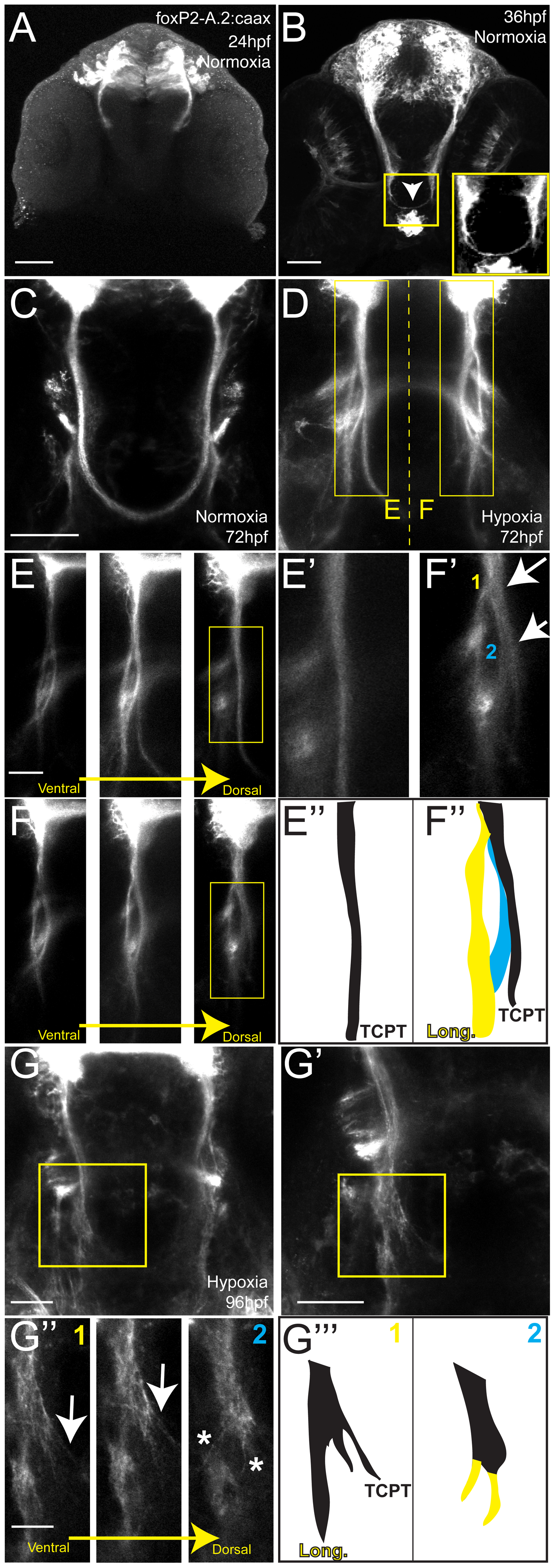
Fig. 2
Hypoxia acts during development of the TCPTc by disrupting axon pathfinding.
Confocal whole-mount images, anti-GFP immunohistochemistry, of Tg(foxP2-A.2:caax) embryos, ventral views, rostral top, except as noted images are maximum intensity projections, scale bars 50 μm except E–E2 and H2 25 μm; H3 12.5 μm. (A) Embryo at 24 hpf, cell bodies and initial axon processes are visible. (B) Embryo at 36 hpf; TCPTc has formed (arrowhead, magnified in inset). (C) TCPTc has formed completely by 72 hpf in normoxia. (D) Disruption of TCPTc in hypoxia. Dotted line shows the subsequent high-resolution pictures from the inset boxes: E–E3 from the left hand TCPTc with normal pathfinding; F–F3 with the disrupted TCPTc. (E) Higher magnification views of embryo at 72 hpf, single confocal slices, showing decussation of TCPT commissural and longitudinal axon tracts at different dorsal-ventral levels. (E2) Inset from dorsal-most level, single confocal slice, shows TCPTc arises as a single axon tract. (E3) Schematic of (E2) showing the single tract that will give rise to the TCPTc. (F) Mirror-images of the disrupted TCPTc from the right-hand of image (D), single confocal slices. The dorsal-most image shows the disrupted axon pathfinding. (F2) High-resolution single confocal slice, shows the two aberrant axon pathfinding errors made, 1 and 2, as axons leave the TCPT to join a longitudinal tract. (F3) Schematic of (F2) illustrating the aberrant pathfinding of axons leaving the TCPTc. (G) Hypoxic embryo at 96 hpf and higher resolution shown in (G2). (G3) is high-resolution ventral-to-dorsal single confocal slices. Arrows point to sparse axons giving rise to the TCPTc. Asterisks demonstrate axons aberrantly turning caudally and failing to join the TCPTc in the dorsal-most image. (G23) Schematic of (G3) illustrating the aberrant pathfinding of axons leaving the TCPTc at different ventral-dorsal levels.