
Fig. S11
- ID
- ZDB-IMAGE-120601-20
- Publication
- Chablais et al., 2012 - The regenerative capacity of the zebrafish heart is dependent on TGFβ signaling
- All Figures
- Figures for Chablais et al., 2012
|
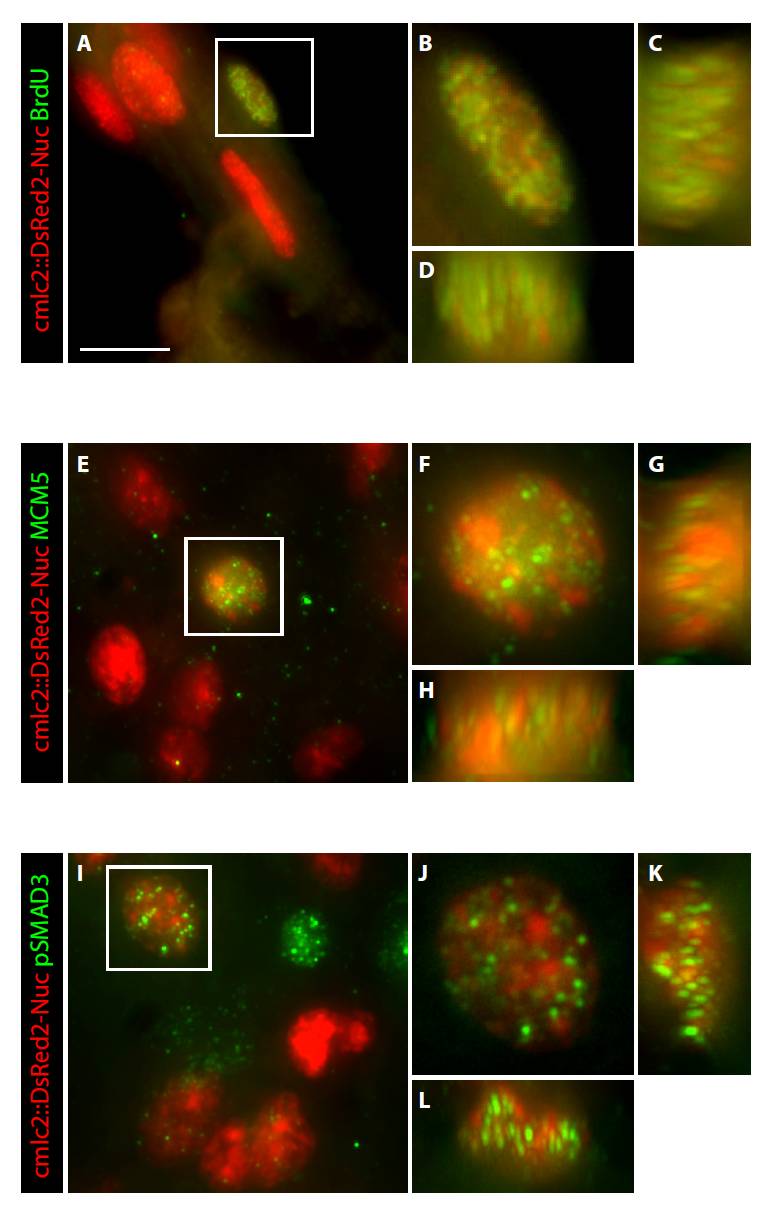
Fig. S11 High-resolution images display co-localization of nuclear markers in the cardiomyocytes of regenerating hearts. (A-L) Heart sections of cmlc2::DsRed2-Nuc transgenic fish were immunostained for BrdU (A-D), Mcm5 (E-H) and pSmad3 (I-L). Some cardiac nuclei that are marked by DsRed2-Nuc expression (red) are co-labeled with the antibody staining (green). The images were acquired using an Olympus IX71 confocal microscope equipped with Photometrics Cool SNAP HQ2 camera. (A-D) Regenerating hearts at 10 dpci after 1 day of BrdU treatment. (E-L) Regenerating hearts at 14 dpci. (B,F,J) A higher magnification of the framed nucleus in the left panels shown as a merged file of 60 optical z-sections. (C,G,K) A z-stack of 60 optical sections along the y-axis of the nucleus that is shown in the adjacent left panels. (D,H,L) A z-stack of 60 optical sections along x-axis of the nucleus that is shown in the upper panels. Scale bar: 10 μm.