
|
Fig. 4
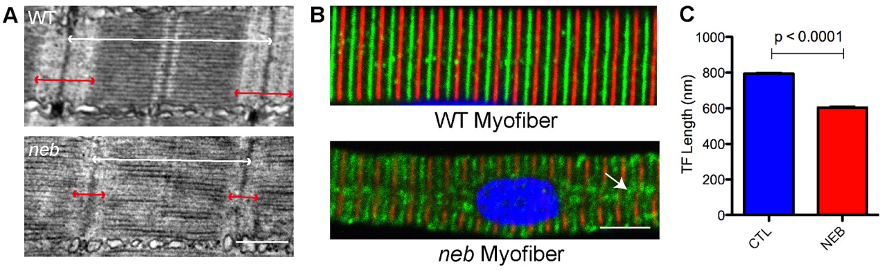
Altered thin filament length in skeletal muscle from neb embryos. (A) Ultrastructural analysis of neb embryos and wild-type clutchmates at 3 dpf. Thin filaments (I bands; marked with red arrows) are qualitatively smaller in neb embryos compared with wild type. Overall sarcomere length is also reduced (white arrows). Scale bar: 500 nm. (B) Co-immunostaining of isolated myofibers with antibodies to α-actinin (red), to mark the Z-lines, and tropomodulin (green), to mark the ends of the thin filaments. Note that α-actinin staining is somewhat diminished in neb embryos and that there are areas of disorganization of tropomodulin staining (arrow). Scale bar: 10 μm. (C) Quantification of thin filament (TF) length. TF length was calculated using electron micrographs by measuring from the Z-line to the beginning of the H zone. TF length in neb zebrafish was significantly reduced (in nm) compared with controls (CTL): 794.3±5.7 vs 603.8±7.9, n=4 averaged measurements, P<0.0001.