
|
Fig. S4
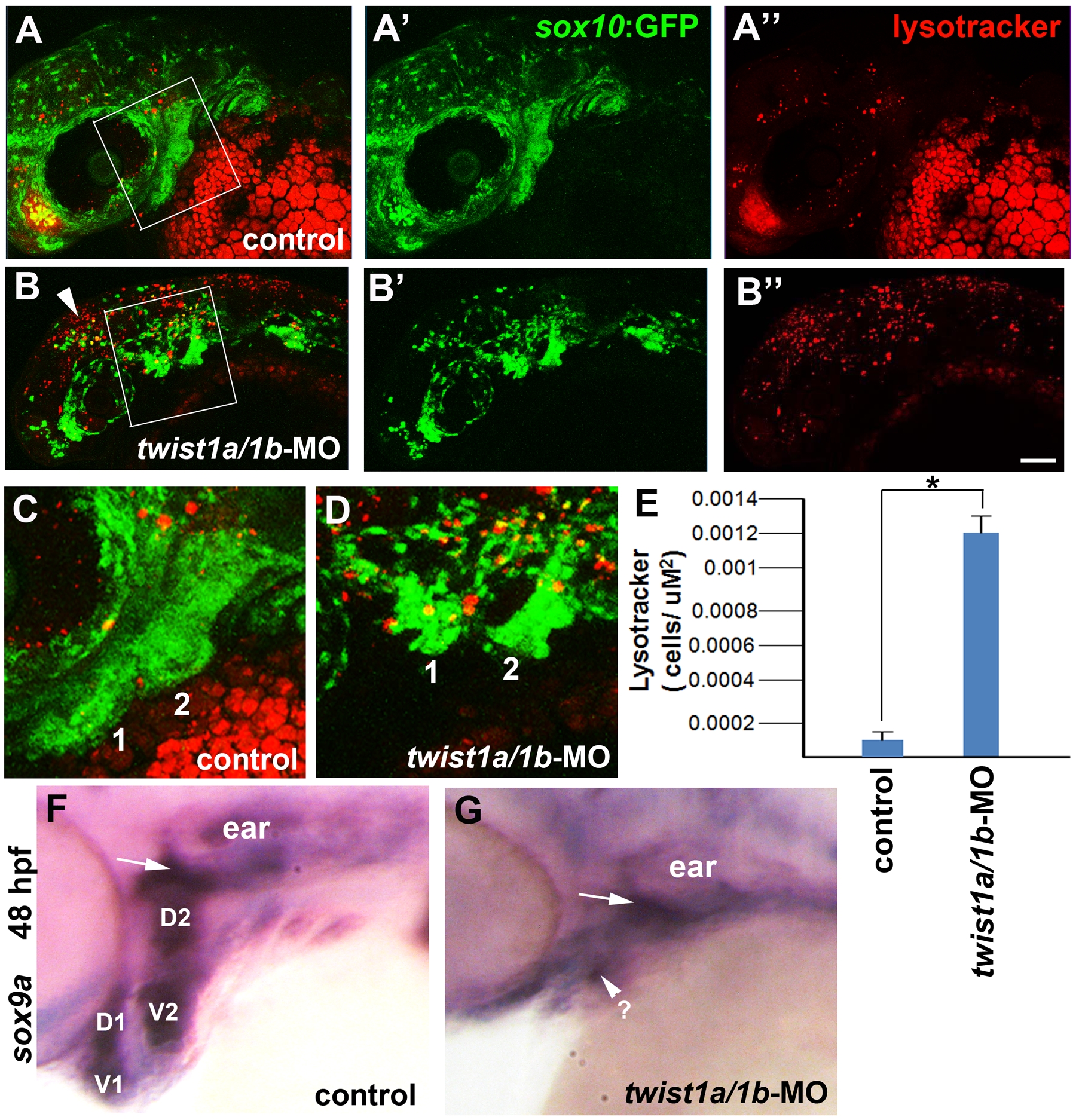
Cell death and sox9a expression in twist1a/1b-MO embryos. (A–D) Confocal projections of Lysotracker Red staining in 36 hpf sox10:GFP transgenic embryos show increased cell death in the pharyngeal arches (shown in high-magnification views in C and D from the boxes in A and B) in twist1a/1b-MO-injected embryos (n = 6) compared to un-injected controls (n = 6). Arrowhead shows increased cell death in more dorsal CNCCs as well. Scale bar = 50 μm. (E) Quantification of Lysotracker-positive cells per arch area. Mandibular and hyoid arches were used for the analysis. Asterisk indicates statistical significance using a Tukey-Kramer HSD test (α = 0.05). (F,G) In situ hybridizations for sox9a at 48 hpf show very reduced expression in the pharyngeal arches of twist1a/1b-MO embryos (n = 5/5) compared to uninjected controls (n = 0/12). Dorsal (D) and ventral (V) pre-chondrogenic domains of the mandibular (1) and hyoid (2) arches are shown for the control. sox9a expression in the pre-chondrogenic domain of the mesoderm-derived otic capsule cartilage (arrows) was less affected in twist1a/1b-MO embryos.