
Image
|
Figure Caption
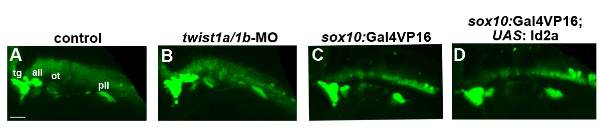
Fig. S3
Cranial ganglionic neurons are unaffected in twist1a/1b-MO and Id2a misexpression embryos. (A–D) Confocal projections of anti-HuC/D immunofluorescence show neurons of the trigeminal (tg), anterior lateral line (all), otic (ot), and posterior lateral line (pll) ganglia at 36 hpf. No major differences in the pattern of anti-HuC/D staining was observed between uninjected control (n = 6), twist1a/1b-MO (n = 6), sox10:Gal4VP16 only control (n = 9), and sox10:Gal4VP16: UAS:Id2a (n = 8) embryos. Scale bar = 50 μm.
Acknowledgments
This image is the copyrighted work of the attributed author or publisher, and
ZFIN has permission only to display this image to its users.
Additional permissions should be obtained from the applicable author or publisher of the image.
Full text @ PLoS Genet.