
|
Fig. 4
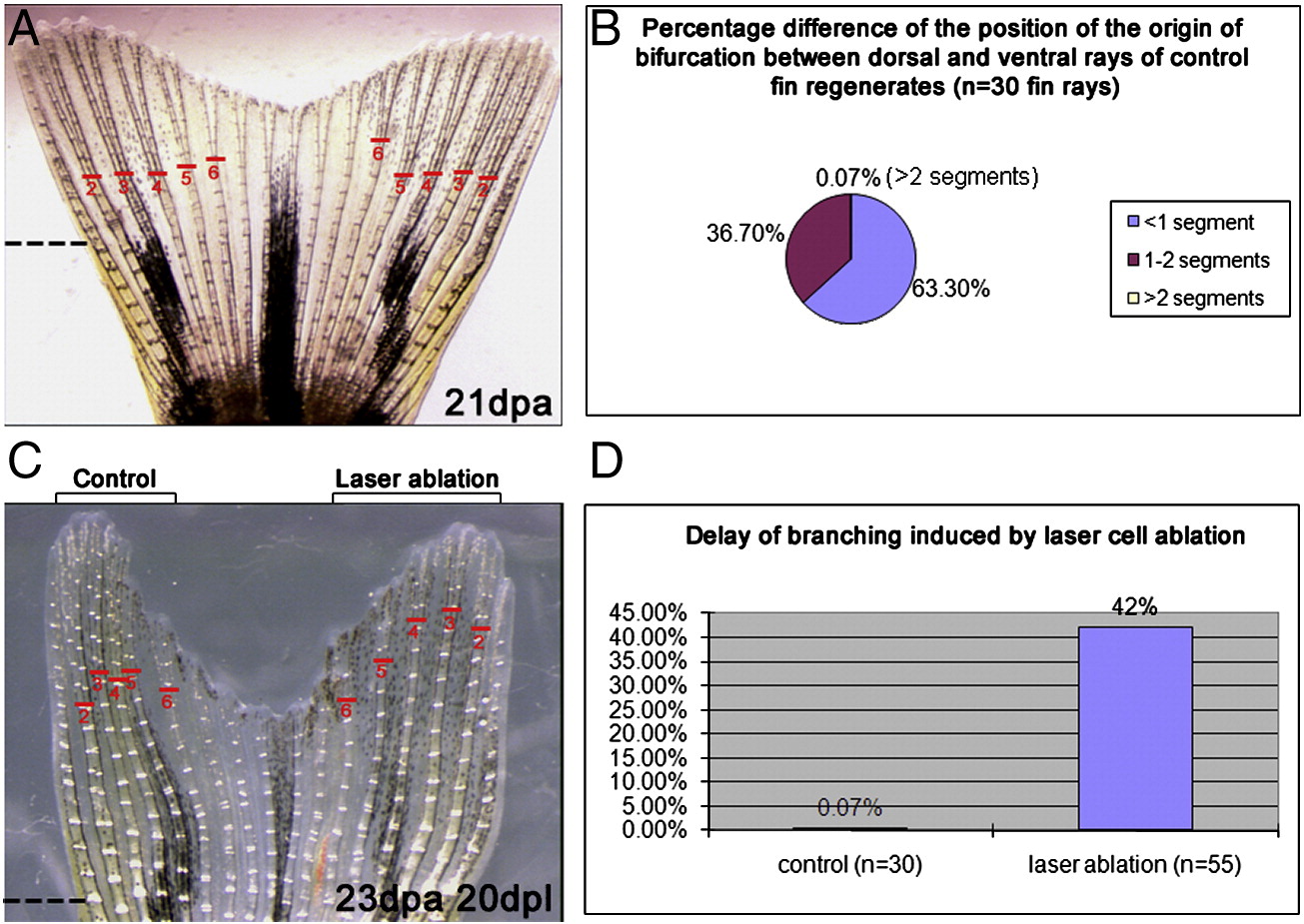
Laser ablation induces a delay of ray branching. (A) Caudal fin regenerate observed at 21 days following amputation below the first branching point of the rays. Ray numbers are shown in red; the corresponding rays on the ventral and dorsal lobes have the same number (ray 1 that never forms bifurcation is not indicated). Red lines indicate the position of the first branching point of five ray regenerates of the ventral and dorsal lobes, respectively. (B) percentage difference in the position of the origin of bifurcation (measured in number of segments) between the corresponding rays of the dorsal and ventral lobes observed in fin regenerates (n = 30) at 21 dpa. Only 0.07% of the rays show a difference greater than 2 segments of the origin of bifurcation. (C) Fin regenerate at 20 dpl of a 2.4shh:gfpABC#15 transgenic fish after laser cell ablation of the GFP positive cells in rays 2, 3, 4, of the ventral lobe at 3 dpa. The red lines indicate the origin of bifurcation in rays 2, 3, 4 in the control side and the side where laser ablation has been performed, respectively. (D) Graph showing the percentage of rays presenting a difference greater than 2 segments in the position of the origin of bifurcation between the ventral and dorsal lobes, in control fins (no ablation) (left bar) and in experimental fins (right bar) in which GFP positive cells have been ablated in rays of the ventral lobe. Dashed lines indicate the level of amputation.
Reprinted from Developmental Biology, 365(2), Zhang, J., Jeradi, S., Strähle, U., and Akimenko, M.A., Laser ablation of the sonic hedgehog-a-expressing cells during fin regeneration affects ray branching morphogenesis, 424-433, Copyright (2012) with permission from Elsevier. Full text @ Dev. Biol.