
|
Fig. 1
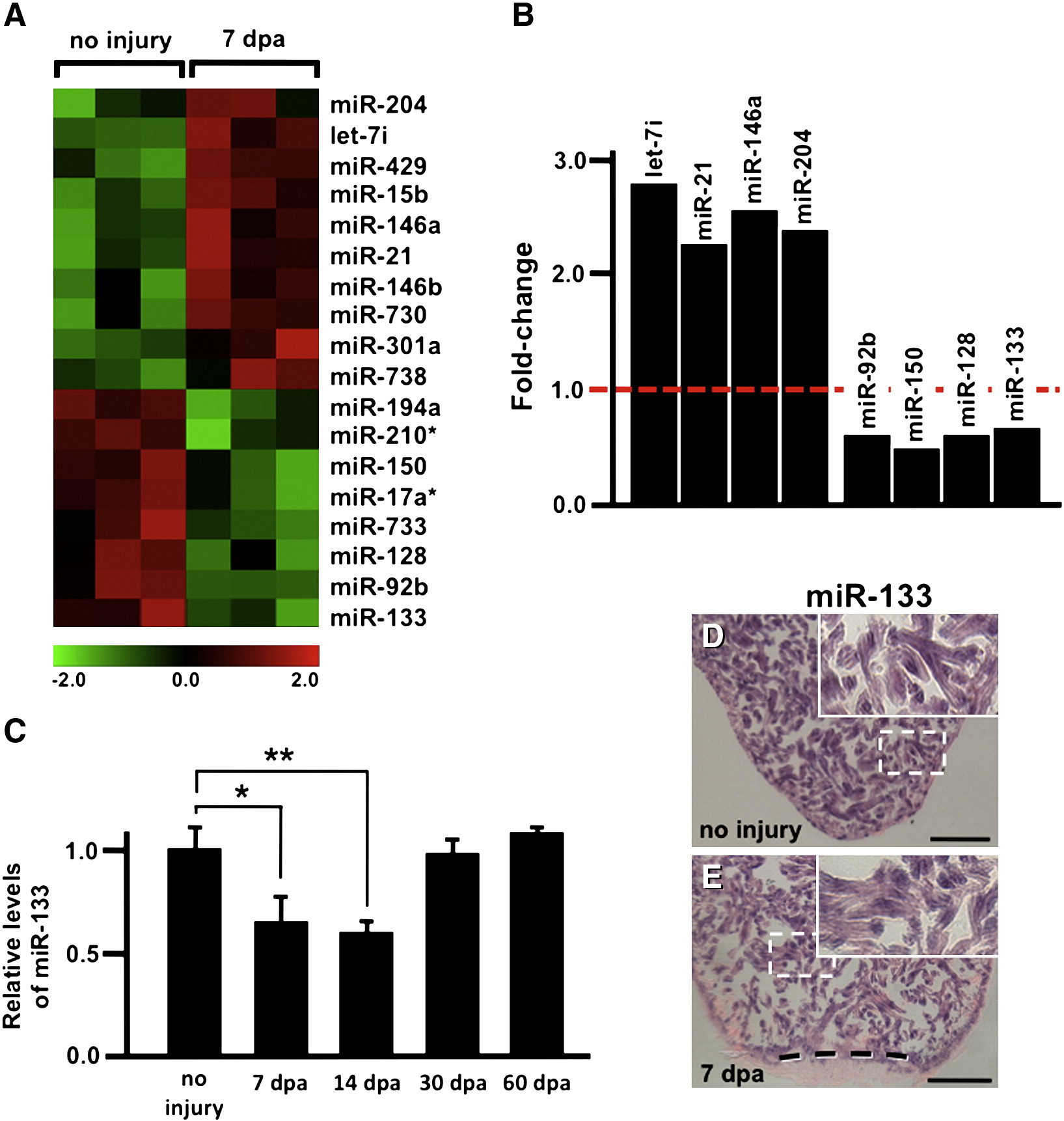
miRNAs are dynamically regulated during myocardial regeneration. A) A heat-map depicts triplicate microarray hybridizations, revealing a subset of miRNAs that are differentially expressed at 7 dpa when compared to uninjured samples. (Green) lower expression; (red) higher expression. B) Real-time quantitative PCR (QPCR) studies confirm the upregulation of let-7i, miR-21, miR-146A and miR-204 and downregulation of miR-92b, miR-150, miR-128 and miR-133 at 7 dpa when compared to uninjured samples. C) QPCR studies show miR-133 levels are high in the uninjured adult heart and reduced at 7 dpa. Levels return to near uninjured levels by 30–60 dpa. D–E) In situ hybridizations reveal miR-133 is restricted to cardiomyocytes under conditions of no injury and at 7 dpa. Error bars in (C) represent SEM, Student′s t-test p-value < 0.05 for * and **; Insets in (D–E), high zoom images of the white dashed rectangle; dashed line in (E) represents approximate amputation plane; dpa, days post-amputation; scale bar in (D–E) represents 100 μm.
Reprinted from Developmental Biology, 365(2), Yin, V.P., Lepilina, A., Smith, A., and Poss, K.D., Regulation of zebrafish heart regeneration by miR-133, 319-327, Copyright (2012) with permission from Elsevier. Full text @ Dev. Biol.