
Image
|
Figure Caption
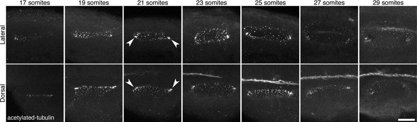
Fig. S1
Presence of cilia in the developing otic vesicle of wild-type zebrafish embryos. Stage series showing cilia, marked by an antibody to acetylated tubulin, in the developing otic vesicle (OV) of wild-type (AB) embryos between 17S and 29S. Arrowheads mark denser ciliated patches at the anterior and posterior poles. All panels are maximum confocal projections through the entire OV. Scale bar: 25 μm.
Acknowledgments
This image is the copyrighted work of the attributed author or publisher, and
ZFIN has permission only to display this image to its users.
Additional permissions should be obtained from the applicable author or publisher of the image.
Full text @ Development