
|
Fig. 5
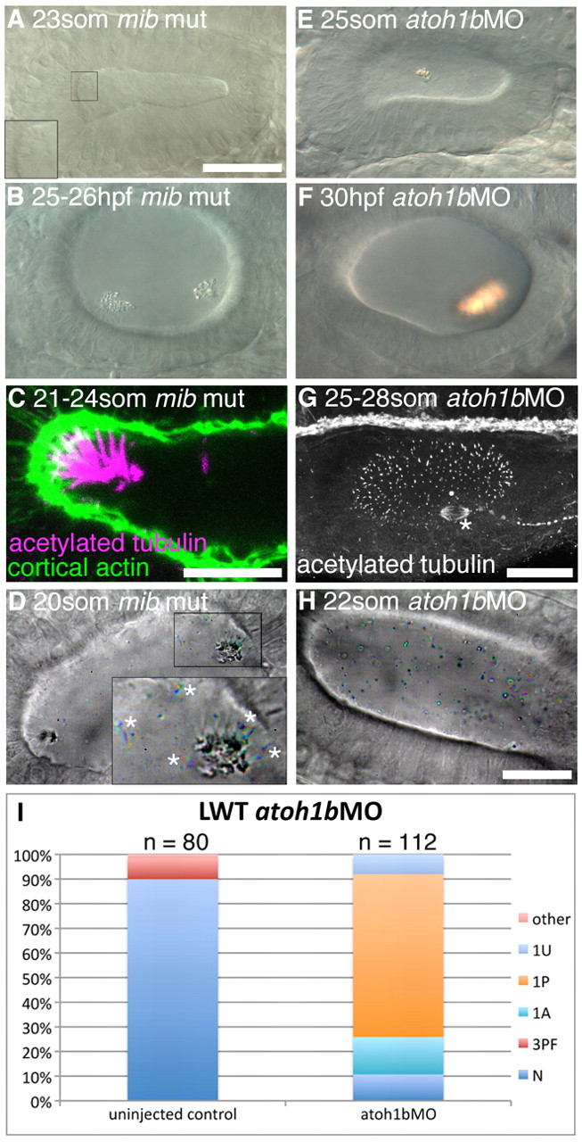
Hair cells are essential for normal otolith formation. (A) Live image of a mib mutant ear at 23S. Inset: ectopic tether cilia. (B) Live image of a 25-26 hpf mib mutant ear. Otoliths do not form correctly. (C) Confocal image showing ectopic tether cilia in a mib mutant ear at 21-24S. (D) Time-to-colour merge of six frames from a high-speed movie of a mib mutant ear. Initially, all ectopic tether cilia nucleate the otolith; motile cilia are present (white asterisks). See supplementary material Movie 12. (E) Live image of atoh1bMO-injected embryo at 25S. Hair cells are absent; an untethered otolith has formed in the middle of the ear. (F) Live image of atoh1bMO-injected embryo at 30 hpf. The single otolith has become tethered to hair cell kinocilia that developed after initial otolith formation. (G) Confocal stack (anti-acetylated tubulin stain) showing presence of non-tether cilia in the ear of an atoh1b MO-injected embryo. The spindle of a dividing cell is visible (asterisk). (H) Time-to-colour merge of six frames of a high-speed movie of an ear from an atoh1b morphant embryo, showing a greater number of untethered otolith precursor particles, and absence of tether cilia and otoliths. Motile cilia are still present (n=3 ears; cf. Fig. 2C). See supplementary material Movie 13. (I) Otolith defects in the ears of atoh1b morphant embryos. N, normal (two otoliths, in correct positions); 1A, one anterior otolith; 1P, one posterior otolith; 1U, one untethered otolith; 3PF, three otoliths, with two posterior otoliths fused; other, other otolith defects. Scale bar: 40 μm in A, for A,B,E,F; 10 μm in C; 25 μm in G; 20 μm; in H, for D,H.