
|
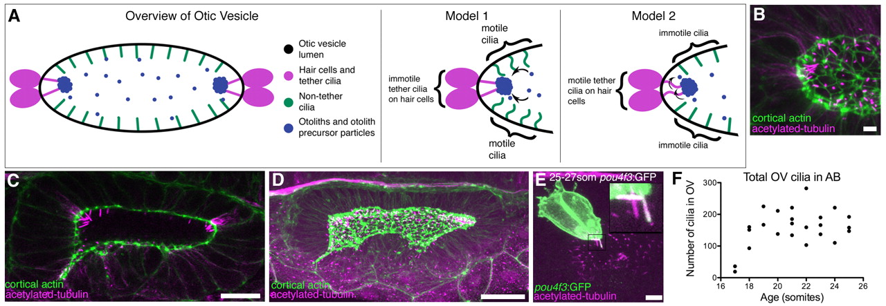
Fig. 1
Presence of cilia in the developing otic vesicle of wild-type zebrafish embryos. (A) Cartoon overview of 18-25S OV and illustrations of current models of otolith formation. (B) Confocal projection showing the anterior OV pole in a 21S embryo (lateral view), stained with FITC-phalloidin (green; cortical actin) and anti-acetylated tubulin (magenta). Scale bar: 5 μm. (C) Single confocal section showing clusters of cilia at the anterior and posterior poles in a 21S stage AB embryo (dorsal view). Scale bar: 20 μm. (D) Confocal projection of the whole OV in a 19-21S LWT embryo. Scale bar: 20 μm. (E) Anti-GFP and anti-acetylated tubulin stain of a Tg(pou4f3:mGFP) embryo. GFP marks the hair cells and their kinocilia. Tether cilia are double-labelled and appear white; other cilia are magenta (inset). Scale bar: 5 μm. (F) Scatter plot showing change in total number of cilia in the OV of AB wild-type embryos.