
|
Fig. 1
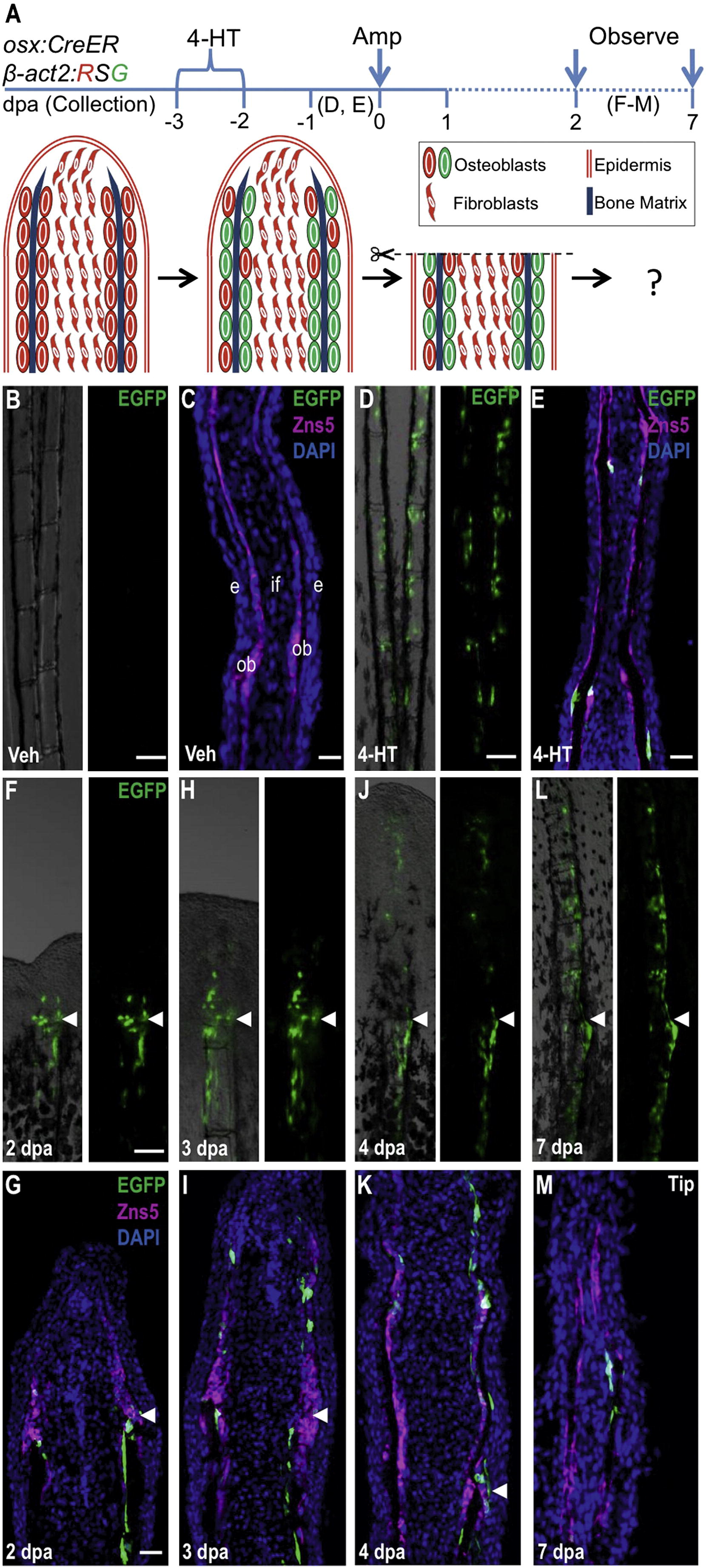
Resident Osteoblasts Contribute New Osteoblasts to Regenerating Fin Structures (A) Cartoon summarizing strategy for inducible, genetic fate mapping of osteoblasts during zebrafish fin regeneration. 4-HT treatment labels osterix-expressing cells with EGFP prior to amputation. (B and C) Uninjured osx:CreER; β-act2:RSG fins, shown as whole mount (B) and in a longitudinal section (C), display no labeling after vehicle treatment. Zns5 (magenta) is an uncharacterized antigen that helps identify osteoblasts lining hemiray bone (Johnson and Weston, 1995). This antibody stains cell membranes and visualizes as noncontiguous staining in sections. (C) The longitudinal fin section is labeled to show structures: intraray fibroblasts (if), osteoblasts (ob), and epidermis (e). (D and E) 4-HT treatment labels many osteoblasts with EGFP in uninjured fins, shown as a whole-mount image (D) and a longitudinal section (E). (F–M) EGFP+ osteoblasts labeled by 4-HT treatment prior to fin amputation contribute labeled progeny to the regenerate, visualized by whole-mount images and in sections at 2 (F and G), 3 (H and I), 4 (J and K), and 7 (L and M) dpa. EGFP fluorescence proximal and distal to the amputation plane is restricted to the osteoblast compartment and is not present in intraray fibroblasts or epidermis. Arrowheads indicate the plane of amputation. Scale bars = 100 μm. See Figure S1.
Reprinted from Developmental Cell, 22(4), Singh, S.P., Holdway, J.E., and Poss, K.D., Regeneration of amputated zebrafish fin rays from de novo osteoblasts, 879-886, Copyright (2012) with permission from Elsevier. Full text @ Dev. Cell